©The Author(s) 2015.
World J Gastroenterol. Apr 7, 2015; 21(13): 4096-4100
Published online Apr 7, 2015. doi: 10.3748/wjg.v21.i13.4096
Published online Apr 7, 2015. doi: 10.3748/wjg.v21.i13.4096
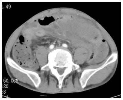
Figure 1 Contrast-enhanced computed tomography of the abdomen indicated segmental dilated small bowel loops with wall thickening and contrast weakening.
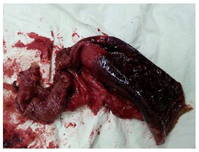
Figure 2 Gross appearance of resected necrotic small bowel.
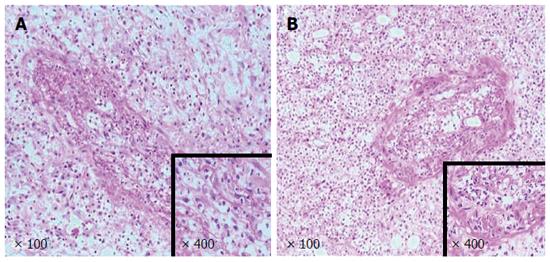
Figure 3 Pathologic findings.
Hematoxylin-eosin staining showed necrosis of the mucosae, congestion, and thrombosis of small vessels in the small bowel (A) and mesentery (B).
Figure 4 Radiologic findings.
Computed tomography showed characteristics of pulmonary infection (A) and hydropericardium (B).
- Citation: Wang QY, Ye XH, Ding J, Wu XK. Segmental small bowel necrosis associated with antiphospholipid syndrome: A case report. World J Gastroenterol 2015; 21(13): 4096-4100
- URL: https://www.wjgnet.com/1007-9327/full/v21/i13/4096.htm
- DOI: https://dx.doi.org/10.3748/wjg.v21.i13.4096


