©The Author(s) 2015.
World J Gastroenterol. Apr 7, 2015; 21(13): 3843-3849
Published online Apr 7, 2015. doi: 10.3748/wjg.v21.i13.3843
Published online Apr 7, 2015. doi: 10.3748/wjg.v21.i13.3843
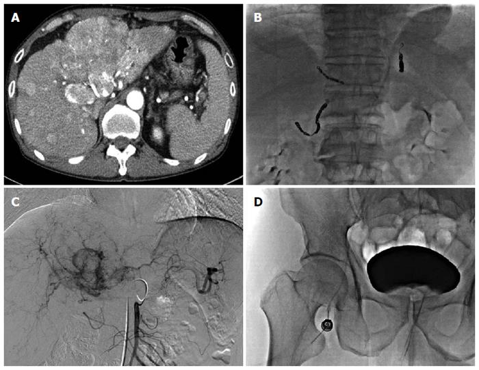
Figure 1 Technical aspects of hepatic artery infusion chemotherapy.
A: Liver dynamic computed tomography showing multinodular hepatocellular carcinoma (HCC) with portal vein thrombosis; B: The embolization of non-target vessels to minimize the flow of chemotherapeutic agents into both uninvolved liver parenchyma and extrahepatic tissues; C: After finding HCC in the feeding artery, the tip of the catheter was located at the proper hepatic or common hepatic artery, chemotherapeutic agents were infused through a pump; D: The proximal end of the catheter was connected to the injection port, which was implanted in a subcutaneous pocket in the right iliac fossa.
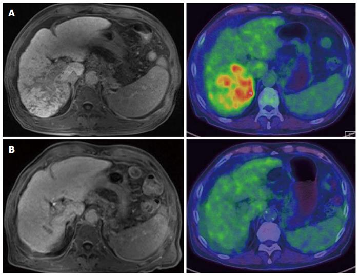
Figure 2 Favorable outcome of patient with infiltrative hepatocellular carcinoma treated by hepatic artery infusion chemotherapy.
A: Patient with infiltrative type hepatocellular carcinoma (HCC) with portal vein thrombosis in liver dynamic magnetic resonance imaging (MRI) showed high FDG uptake; B: After hepatic artery infusion chemotherapy, this patient showed no viable HCC except focal portal vein thrombosis in a follow-up liver MRI and positron emission tomography/computed tomography.
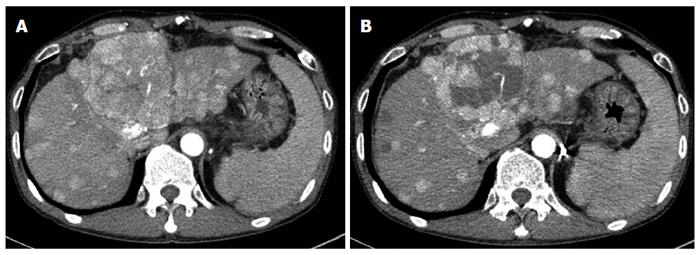
Figure 3 Favorable response of patient with multinodular hepatocellular carcinoma treated by hepatic artery infusion chemotherapy.
A: Patient with multinodular type hepatocellular carcinoma (HCC) with portal vein thrombosis in baseline liver dynamic computed tomography (CT); B: After hepatic artery infusion chemotherapy, this patient showed partial necrosis of HCC in a follow-up liver dynamic CT.
- Citation: Song MJ. Hepatic artery infusion chemotherapy for advanced hepatocellular carcinoma. World J Gastroenterol 2015; 21(13): 3843-3849
- URL: https://www.wjgnet.com/1007-9327/full/v21/i13/3843.htm
- DOI: https://dx.doi.org/10.3748/wjg.v21.i13.3843


