
















©The Author(s) 2015.
World J Gastroenterol. Mar 21, 2015; 21(11): 3425-3428
Published online Mar 21, 2015. doi: 10.3748/wjg.v21.i11.3425
Published online Mar 21, 2015. doi: 10.3748/wjg.v21.i11.3425
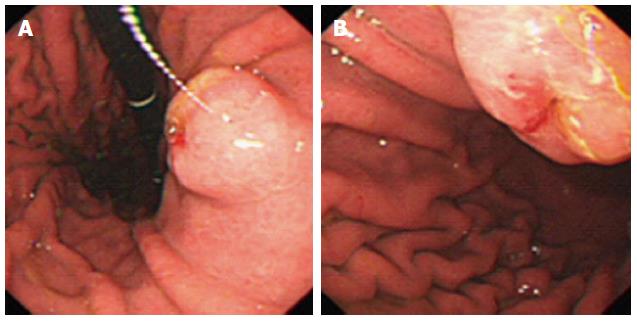
Figure 1 Electronic gastroscopy showed a papillary mass in lesser curvature of the stomach, and some bile on the surface of the mass.
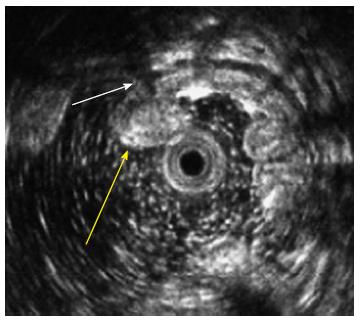
Figure 2 Endoscopic ultrasonography revealed a homogeneously hyperechoic submucosal mass (yellow arrow) with clear margin and a hypoechoic tunnel inside (white arrow).
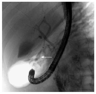
Figure 3 Endoscopic retrograde cholangiopancreatography showed a tubular structure (white arrow) between left hepatic lobe and lesser curvature of the stomach.
GB: Gallbladder.
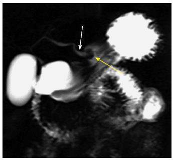
Figure 4 Magnetic resonance cholangiopancreatography revealed the variant bile duct (white arrow) originating from left hepatic duct drained into the lesser curvature of stomach.
Note the hyperintense tubular structure (yellow arrow).
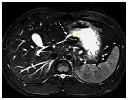
Figure 5 Axial T2WI, a tubular structure (white arrow) with hyperintense signal and its opening (yellow arrow) in stomach can be found as well.
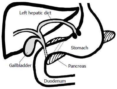
Figure 6 Hand drawing of this case better shows the relationship between variant bile duct and the lesser curvature of the stomach.
- Citation: Guan J, Zhang L, Chu JP, Lin SC, Li ZP. Congenital left intrahepatic bile duct draining into gastric wall mimicking biliary reflux gastritis. World J Gastroenterol 2015; 21(11): 3425-3428
- URL: https://www.wjgnet.com/1007-9327/full/v21/i11/3425.htm
- DOI: https://dx.doi.org/10.3748/wjg.v21.i11.3425