©The Author(s) 2015.
World J Gastroenterol. Mar 21, 2015; 21(11): 3420-3424
Published online Mar 21, 2015. doi: 10.3748/wjg.v21.i11.3420
Published online Mar 21, 2015. doi: 10.3748/wjg.v21.i11.3420
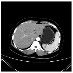
Figure 1 Enhancing computed tomography confirmed a hypo-dense ellipse occupying the upper pole of the spleen.
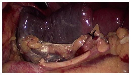
Figure 2 After ligating the upper branches of the splenic artery, an ischemic demarcation line on the splenic surface is pronounced.
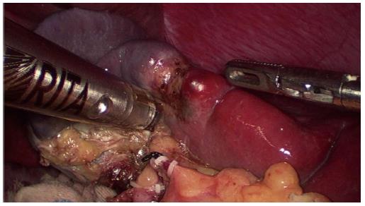
Figure 3 HabibTM 4X was inserted into the splenic parenchyma along the well-defined ischemic demarcation line for coagulating and sealing blood vessels.
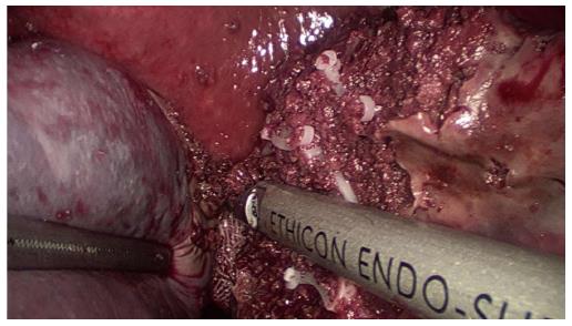
Figure 4 Splenic parenchyma was divided bloodlessly with an Ultracision Harmonic Scalpel.
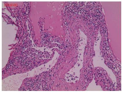
Figure 5 Pathological examination of the splenic lymphangioma.
HE staining, original magnification × 40.
- Citation: Wang WD, Lin J, Wu ZQ, Liu QB, Ma J, Chen XW. Partial splenectomy using a laparoscopic bipolar radiofrequency device: A case report. World J Gastroenterol 2015; 21(11): 3420-3424
- URL: https://www.wjgnet.com/1007-9327/full/v21/i11/3420.htm
- DOI: https://dx.doi.org/10.3748/wjg.v21.i11.3420


