©The Author(s) 2015.
World J Gastroenterol. Mar 14, 2015; 21(10): 3150-3153
Published online Mar 14, 2015. doi: 10.3748/wjg.v21.i10.3150
Published online Mar 14, 2015. doi: 10.3748/wjg.v21.i10.3150
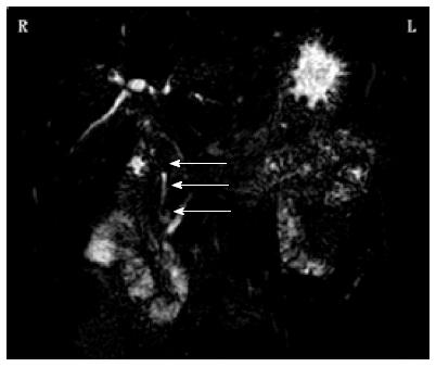
Figure 1 Magnetic resonance cholangiopancreatography shows numerous masses in the common bile duct.
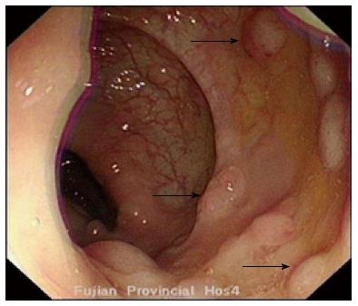
Figure 2 Colonoscopy shows multiple polyps in the colon.
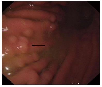
Figure 3 Gastroscopy shows multiple polyps in the duodenum and the distal stomach.
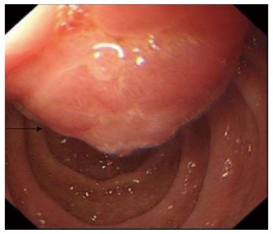
Figure 4 Ampulla of Vater is apparently protuberant and the surface is rough.
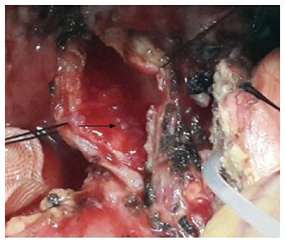
Figure 5 Numerous polyps fill the middle and distal common bile duct.
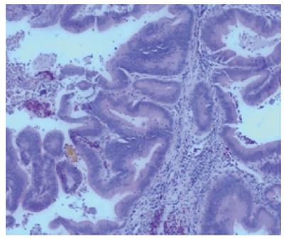
Figure 6 Common bile duct masses are tubular adenomas (HE, × 10).
- Citation: Yan ML, Pan JY, Bai YN, Lai ZD, Chen Z, Wang YD. Adenomas of the common bile duct in familial adenomatous polyposis. World J Gastroenterol 2015; 21(10): 3150-3153
- URL: https://www.wjgnet.com/1007-9327/full/v21/i10/3150.htm
- DOI: https://dx.doi.org/10.3748/wjg.v21.i10.3150


