©The Author(s) 2015.
World J Gastroenterol. Mar 14, 2015; 21(10): 2982-2987
Published online Mar 14, 2015. doi: 10.3748/wjg.v21.i10.2982
Published online Mar 14, 2015. doi: 10.3748/wjg.v21.i10.2982
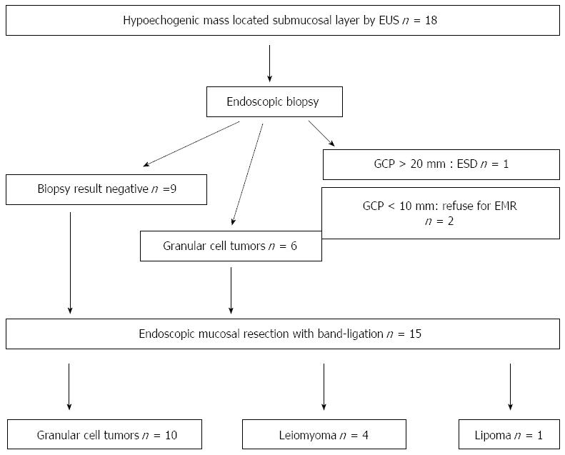
Figure 1 Flow chart of endoscopic treatment for esophageal tumor located in the submucosal layer by endoscopic ultrasonography.
EUS: Endoscopic ultrasonography; GCP: Granular cell tumor; EMR: Endoscopic mucosal resection; ESD: Endoscopic submucosal dissection.
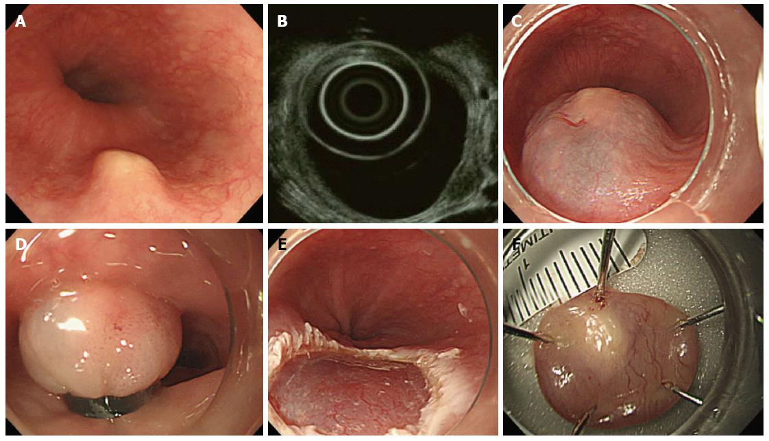
Figure 2 Endoscopic mucosal resection using band-ligation.
A: Endoscopic view of esophageal granular cell tumor; B: Endoscopic ultrasound showed a hypoechogenic lesion in the submucosal layer; C: Submucosal injection was performed; D: The tumor was ligated with the elastic band after submucosal solution injection; E: Ulcer after resection; F: Resected specimen.
- Citation: Hong JB, Choi CW, Kim HW, Kang DH, Park SB, Kim SJ, Kim DJ. Endoscopic resection using band ligation for esophageal SMT in less than 10 mm. World J Gastroenterol 2015; 21(10): 2982-2987
- URL: https://www.wjgnet.com/1007-9327/full/v21/i10/2982.htm
- DOI: https://dx.doi.org/10.3748/wjg.v21.i10.2982


