©The Author(s) 2015.
World J Gastroenterol. Mar 14, 2015; 21(10): 2896-2904
Published online Mar 14, 2015. doi: 10.3748/wjg.v21.i10.2896
Published online Mar 14, 2015. doi: 10.3748/wjg.v21.i10.2896
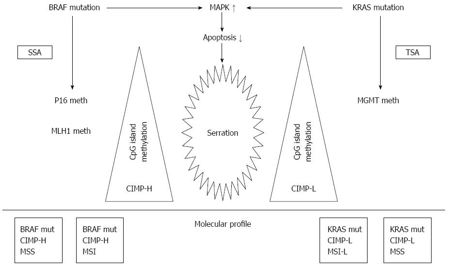
Figure 1 Schematic representation of the sessile and traditional serrated pathways[18].
MSI: Microsatellites instability; CIMP-H, CIMP-L: CpG island methylation promotor-high, CIMP-bas; MLH1: MutL homolog 1; MGMT: MethylGuanine DNA methyltransferase; SSA: Sessile serrated adenomas.
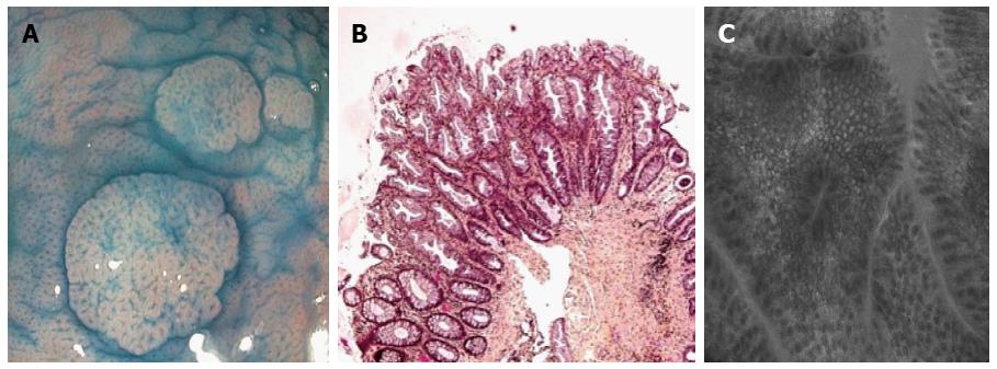
Figure 2 Hyperplastic polyp in chromoendoscopy (A), histology (B) and confocal endomicroscopy (C).
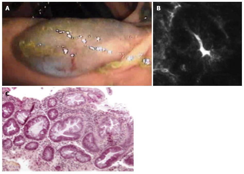
Figure 3 Sessile serrated adenoma in chromoendoscopy (A), confocal endomicroscopy (B) and histology (C)
Figure 4 Traditional serrated polyp in chromoendoscopy (A), confocal endomicroscopy (B) and histology (C).
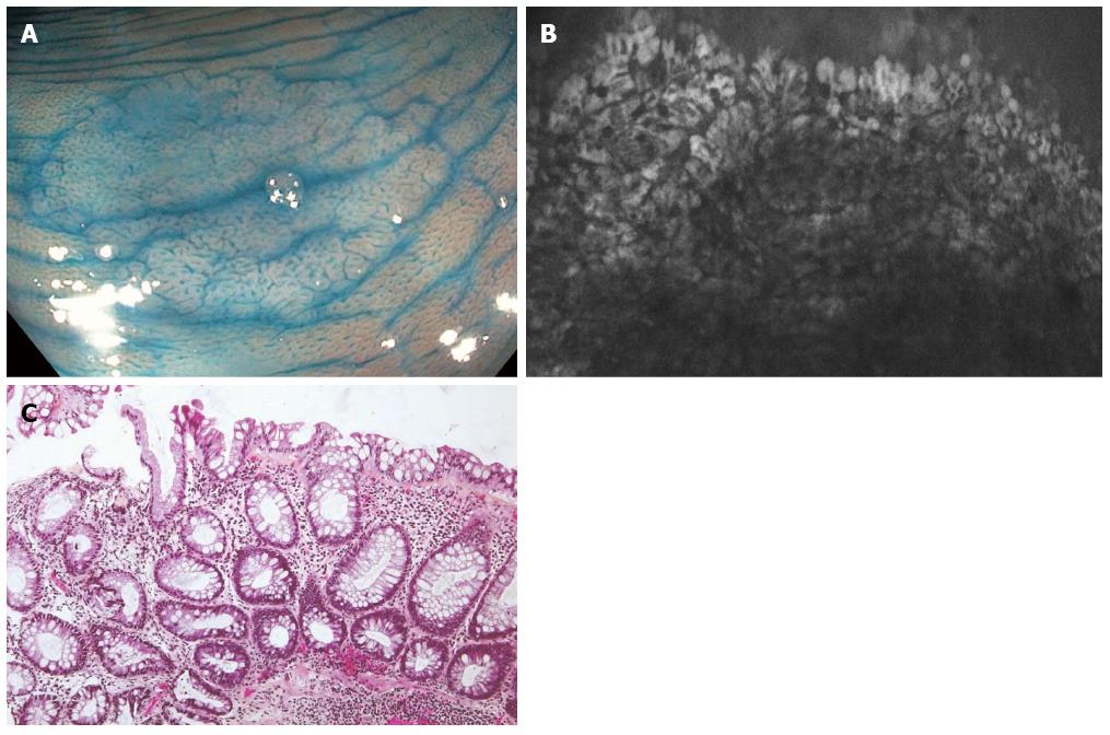
Figure 5 Endoscopic (A), endomicroscopic (B) and histologic (C) characteristics of serrated lesions in patients with inflammatory bowel disease.
- Citation: Moussata D, Boschetti G, Chauvenet M, Stroeymeyt K, Nancey S, Berger F, Lecomte T, Flourié B. Endoscopic and histologic characteristics of serrated lesions. World J Gastroenterol 2015; 21(10): 2896-2904
- URL: https://www.wjgnet.com/1007-9327/full/v21/i10/2896.htm
- DOI: https://dx.doi.org/10.3748/wjg.v21.i10.2896


