©2014 Baishideng Publishing Group Co.
World J Gastroenterol. Feb 21, 2014; 20(7): 1882-1886
Published online Feb 21, 2014. doi: 10.3748/wjg.v20.i7.1882
Published online Feb 21, 2014. doi: 10.3748/wjg.v20.i7.1882
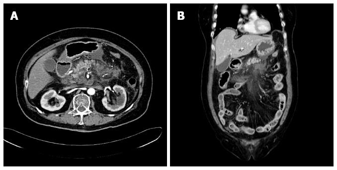
Figure 1 Fatty infiltration and fluid accumulation around the peripancreatic area observed by computerized tomography.
Computerized tomography scans taken upon the patient’s admission to the hospital. A: Axial view; B: Coronal view.
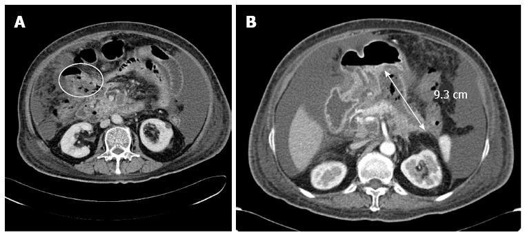
Figure 2 A fistula and a pseudocyst identified through computerized tomography.
A: There was a fistula in the right side of the colon (yellow circle); B: There was a pseudocyst approximately 9.3 cm long (demarcated by double-headed arrow) that contained air.
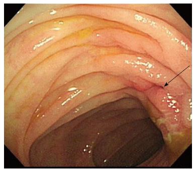
Figure 3 Fistula opening identified by colonoscopy.
There was a fistular opening (arrow) that surrounded edematous and hyperemic mucosa at the hepatic flexure.
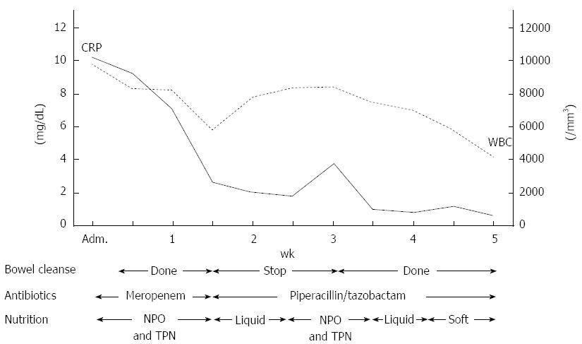
Figure 4 Timetable of bowel cleansing, administration of antibiotics and nutrition.
After daily bowel cleansing using a polyethylene glycol solution, white blood cell (WBC) count and C-reactive protein (CRP) value were decreased. After the initial bowel cleanse was stopped, we observed an elevated WBC count and CRP level. The bowel cleanse was continued until 6 wk after admission. Adm: Admission; NPO: Nil per os; TPN: Total parenteral nutrition.
- Citation: Kwon JC, Kim BY, Kim AL, Kim TH, Park MI, Jung HJ, Lim JH, Jung JK, Kim HS, Lee DW. Pancreatic pseudocystocolonic fistula treated without surgical or endoscopic intervention. World J Gastroenterol 2014; 20(7): 1882-1886
- URL: https://www.wjgnet.com/1007-9327/full/v20/i7/1882.htm
- DOI: https://dx.doi.org/10.3748/wjg.v20.i7.1882


