Copyright
©2014 Baishideng Publishing Group Co.
World J Gastroenterol. Feb 14, 2014; 20(6): 1402-1423
Published online Feb 14, 2014. doi: 10.3748/wjg.v20.i6.1402
Published online Feb 14, 2014. doi: 10.3748/wjg.v20.i6.1402
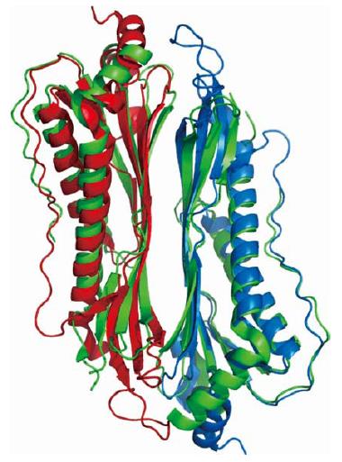
Figure 1 Cartoon view of the superimposed Cα traces of HpaA paralogs.
HP0410 dimer (3bgh, green) and HP0492 dimer (2i9i, red and light blue). Cα root mean square deviation between equivalent atoms corresponds to 2.4 Å.
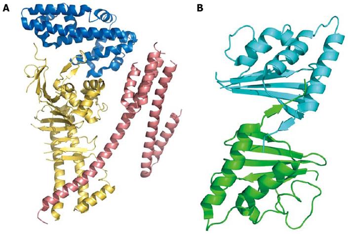
Figure 2 Cartoon model of Cag proteins.
A: The N-terminal portion of cytotoxic-associated genes, CagA, the effector protein injected into the host cell through type IV secretion system. The three domains (residues 24-221, blue; 303-644, yellow; 645-824, salmon; coordinates from PDB 4DVY) are shown in different colors; B: CagD dimer. The two monomers are linked together by a disulfide bridge between the two C-terminal β-strands. Coordinates from PDB 3CWX.
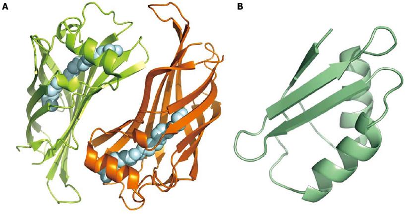
Figure 3 Binding and transport proteins.
A: Cartoon model of HP1286 lipocalin dimer. The two monomers, related by a two-fold axis, bind in the inner cavity a molecule of erucamide (silver spheres; PDB 3HPE); B: Nuclear magnetic resonance structure of apo-CopP, a copper binding regulatory protein of 66 amino acid residues (PDB 1YG0).
Figure 4 Toxins.
A: Cartoon model of the p55 domain of vacuolating toxin (VacA) (coordinates from PDB 2QV3). The structure is a predominantly right-handed parallel β-helix, and the domain mediates the binding of VacA to the host cell; B: Cartoon model of a truncated form of tumor-necrosis-factor α (TNFα) inducing protein, a virulence factor that enters gastric cells and stimulates both the production of TNFα and the nuclear factor kappa B pathway (coordinates PDB 2WCR).
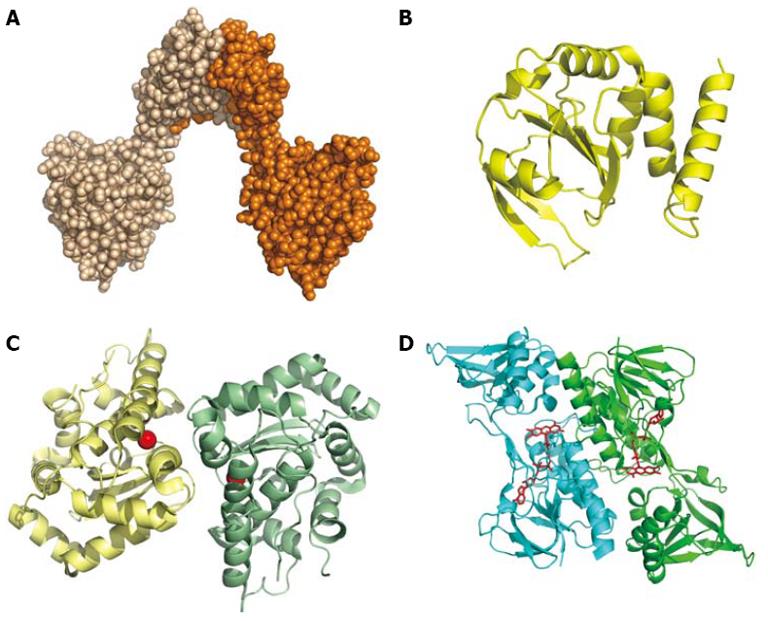
Figure 5 Redox proteins.
A: Space-filling model of the dimer of DsbG (HP0231; PDB 3TDG); B: Cartoon of DsbC (HP0377; Coordinates PDB 4FYC), an enzyme with a thioredoxin-like fold possibly involved in cytochrome c assembly; C: Cartoon of the dimeric Fe-superoxide dismutase (Coordinates PDB 3CEI). The iron ion is represented by a red sphere; D: Dimeric thioredoxin reductase (Coordinates PDB 3ISH). The FAD bound is shown as a ball-and-stick model.
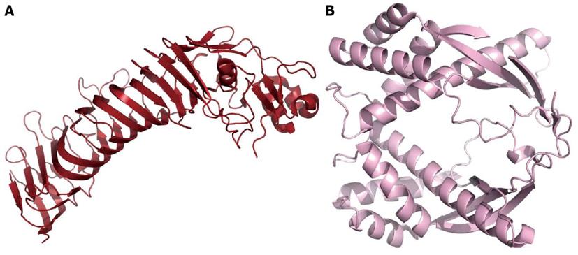
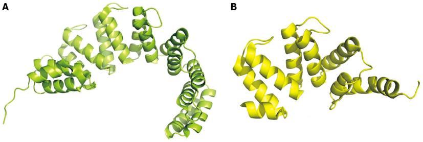
Figure 6 Two examples of solenoid-class proteins.
A: HcpB (HP0336, coordinates PDB1KLX); B: HcpC (HP1098, coordinates PDB 1OUV).
-
Citation: Zanotti G, Cendron L. Structural and functional aspects of the
Helicobacter pylori secretome. World J Gastroenterol 2014; 20(6): 1402-1423 - URL: https://www.wjgnet.com/1007-9327/full/v20/i6/1402.htm
- DOI: https://dx.doi.org/10.3748/wjg.v20.i6.1402


