©2014 Baishideng Publishing Group Co.
World J Gastroenterol. Feb 7, 2014; 20(5): 1259-1267
Published online Feb 7, 2014. doi: 10.3748/wjg.v20.i5.1259
Published online Feb 7, 2014. doi: 10.3748/wjg.v20.i5.1259
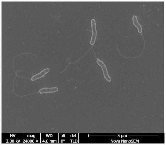
Figure 1 Electron microscopic image of Campylobacter concisus.
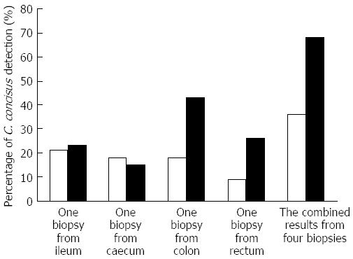
Figure 2 An example showing that collection of multiple intestinal biopsies increases the detection of intestinal prevalence of Campylobacter concisus.
Data included in Figure 2 were from reference 20. In both patients with inflammatory bowel disease (IBD) (right column) and healthy controls (left column), the detection of intestinal prevalence of Campylobacter concisus (C. concisus) was greatly increased using four biopsies as compared to using one biopsy. In healthy controls, C. concisus detection rates in a single biopsy collected from ileum, caecum, colon and rectum were 21%, 18%, 18% and 9% respectively. If results from the four biopsies were used in determining the intestinal prevalence of C. concisus, the intestinal prevalence of C. concisus in healthy controls was 36%. Similarly, in patients with IBD, C. concisus detection rates in a single biopsy collected from ileum, caecum, colon and rectum were 23%, 15%, 43% and 26% respectively, and the intestinal prevalence of C. concisus in patients with IBD was 68% when four biopsies were taken into consideration.
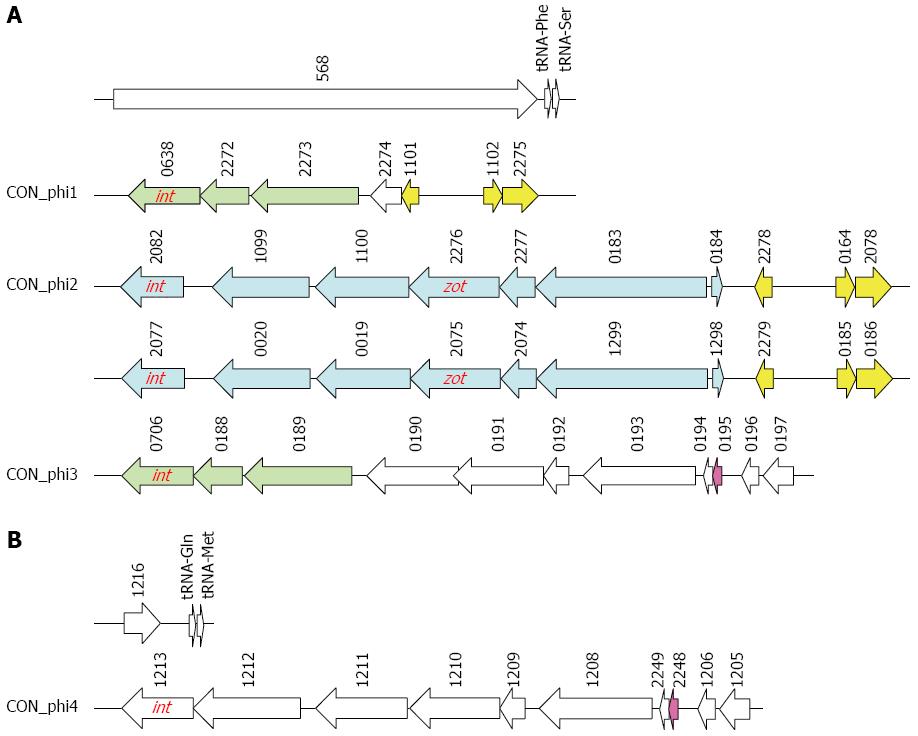
Figure 3 Genetic structures of putative prophage identified in Campylobacter concisus strain 13826.
A: Multiple prophages at nucleotide position between 1576686 and 1614075; B: One putative prophage at nucleotide position between 939158 and 946901. Identical genes were indicated with the same color. int: A gene encodes phage integrase; zot: Zonula occludens toxin gene. The numbers are the locus tags.
-
Citation: Zhang L, Lee H, Grimm MC, Riordan SM, Day AS, Lemberg DA.
Campylobacter concisus and inflammatory bowel disease. World J Gastroenterol 2014; 20(5): 1259-1267 - URL: https://www.wjgnet.com/1007-9327/full/v20/i5/1259.htm
- DOI: https://dx.doi.org/10.3748/wjg.v20.i5.1259


