Copyright
©2014 Baishideng Publishing Group Inc.
World J Gastroenterol. Dec 7, 2014; 20(45): 17171-17178
Published online Dec 7, 2014. doi: 10.3748/wjg.v20.i45.17171
Published online Dec 7, 2014. doi: 10.3748/wjg.v20.i45.17171
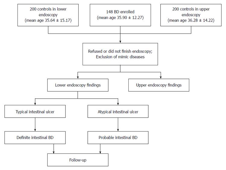
Figure 1 Study protocol and algorithm for the diagnosis of intestinal Behcet’s disease.
Complete, incomplete, and suspected subtypes of systemic Behcet’s disease (BD) were classified according to the diagnostic criteria of the Research Committee of Japan 1987.
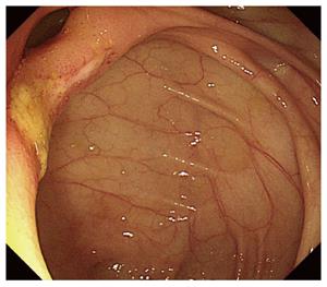
Figure 2 Ileocolonoscopic appearance of patient 10 with an ulcer in her ileocecal valve.
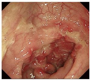
Figure 3 Colonoscopic appearance of patient 2 with ulcers in his transverse colon.
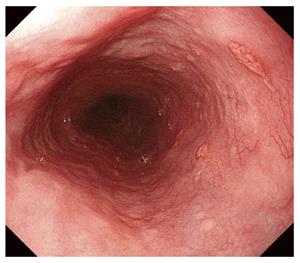
Figure 4 Endoscopic appearance of patient 2 with ulcers in his esophagus.
- Citation: Zou J, Shen Y, Ji DN, Zheng SB, Guan JL. Endoscopic findings of gastrointestinal involvement in Chinese patients with Behcet’s disease. World J Gastroenterol 2014; 20(45): 17171-17178
- URL: https://www.wjgnet.com/1007-9327/full/v20/i45/17171.htm
- DOI: https://dx.doi.org/10.3748/wjg.v20.i45.17171


