Copyright
©2014 Baishideng Publishing Group Inc.
World J Gastroenterol. Dec 7, 2014; 20(45): 17148-17154
Published online Dec 7, 2014. doi: 10.3748/wjg.v20.i45.17148
Published online Dec 7, 2014. doi: 10.3748/wjg.v20.i45.17148
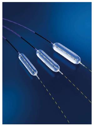
Figure 1 Endoscopic papillary large balloon dilation: Controlled Radial Expansion 12-20 mm wire-guided type balloon 5.
5 cm (Boston Scientific Corp., Natick, MA).
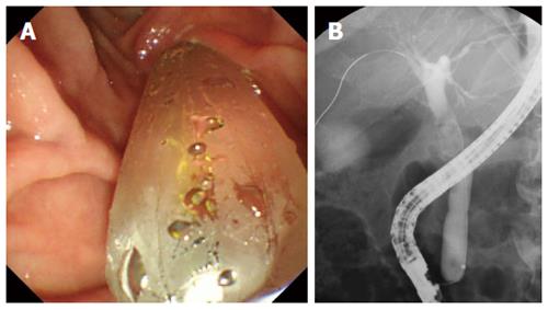
Figure 2 View of papilla gradually dilated.
A: The papilla was gradually dilated using a large balloon. Dilation was continued until the notch on the balloon disappeared (endoscopic image); B: The papilla was gradually dilated until the notch on the balloon disappeared (fluoroscopic image).
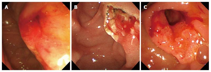
Figure 3 Duodenal papilla after endoscopic procedure.
A: Papillary balloon dilation; B: The duodenal papilla after endoscopic sphincterotomy; C: The duodenal papilla after endoscopic papillary large balloon dilation.
- Citation: Sakai Y, Tsuyuguchi T, Kawaguchi Y, Hirata N, Nakaji S, Kitamura K, Mikami S, Fujimoto T, Ijima M, Kurihara E, Oana S, Nishino T, Tamura R, Sakamoto D, Nakamura M, Nishikawa T, Sugiyama H, Yoshida H, Mine T, Yokosuka O. Endoscopic papillary large balloon dilation for removal of bile duct stones. World J Gastroenterol 2014; 20(45): 17148-17154
- URL: https://www.wjgnet.com/1007-9327/full/v20/i45/17148.htm
- DOI: https://dx.doi.org/10.3748/wjg.v20.i45.17148


