©2014 Baishideng Publishing Group Inc.
World J Gastroenterol. Oct 21, 2014; 20(39): 14510-14514
Published online Oct 21, 2014. doi: 10.3748/wjg.v20.i39.14510
Published online Oct 21, 2014. doi: 10.3748/wjg.v20.i39.14510
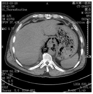
Figure 1 Computed tomography scan of the anacardium revealing posterior mediastinal widening, hydrops, and pneumatosis, mimicking findings of esophageal hiatus hernia.
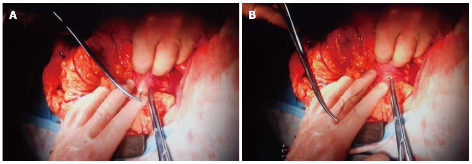
Figure 2 Stomach surgery for the removal of the hard thick fishbone (A and B).
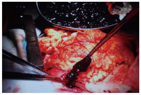
Figure 3 Removal of a massive blood clot from the stomach.
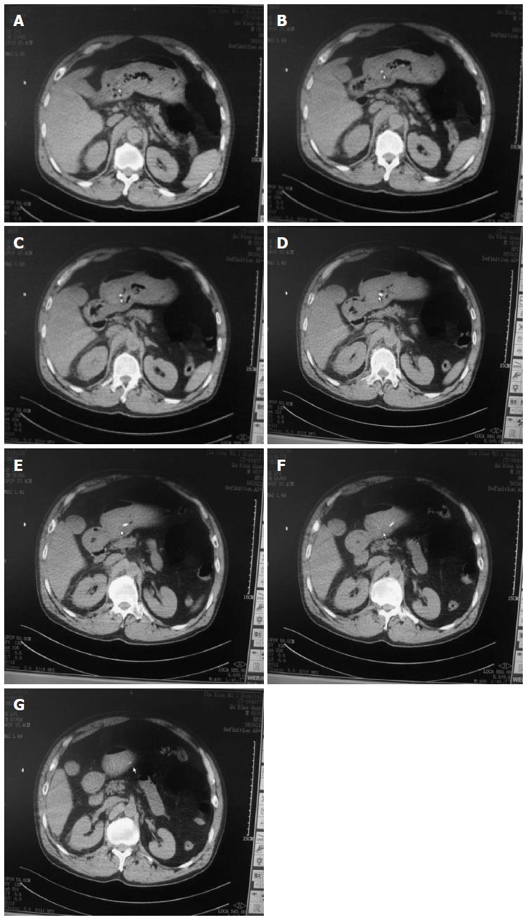
Figure 4 Computed tomography scan of the abdomen revealing the fishbone in the stomach that penetrated 7 layers of the stomach wall (A-G).
- Citation: Lu YP, Yao M, Zhou XY, Huang B, Qi WB, Chen ZH, Xu LS. False esophageal hiatus hernia caused by a foreign body: A fatal event. World J Gastroenterol 2014; 20(39): 14510-14514
- URL: https://www.wjgnet.com/1007-9327/full/v20/i39/14510.htm
- DOI: https://dx.doi.org/10.3748/wjg.v20.i39.14510


