©2014 Baishideng Publishing Group Inc.
World J Gastroenterol. Oct 21, 2014; 20(39): 14301-14307
Published online Oct 21, 2014. doi: 10.3748/wjg.v20.i39.14301
Published online Oct 21, 2014. doi: 10.3748/wjg.v20.i39.14301
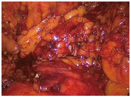
Figure 1 Standard right hemicolectomy.
Completed standard dissection of a right hemicolectomy with ligated ileocolic pedicle (A) and right branch of the middle colic artery (B). Note no dissection over the superior mesenteric vein or artery.
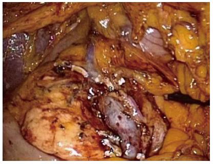
Figure 2 Complete mesocolic excision right hemicolectomy.
complete mesocolic excision dissection of a right hemicolectomy showing the superior mesenteric vein and artery and ligated Ileocolic (A) and right branch of the middle colic (B) pedicles.
- Citation: Chow CFK, Kim SH. Laparoscopic complete mesocolic excision: West meets East. World J Gastroenterol 2014; 20(39): 14301-14307
- URL: https://www.wjgnet.com/1007-9327/full/v20/i39/14301.htm
- DOI: https://dx.doi.org/10.3748/wjg.v20.i39.14301


