Copyright
©2014 Baishideng Publishing Group Inc.
World J Gastroenterol. Oct 14, 2014; 20(38): 13899-13903
Published online Oct 14, 2014. doi: 10.3748/wjg.v20.i38.13899
Published online Oct 14, 2014. doi: 10.3748/wjg.v20.i38.13899
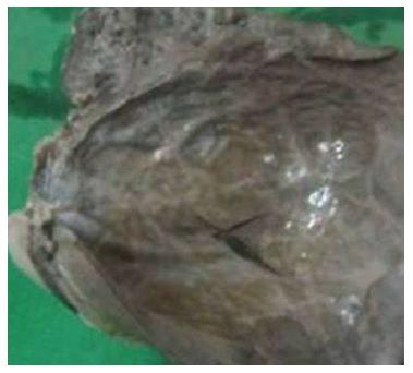
Figure 1 Huge primary splenic cyst with glistening smooth inner wall.
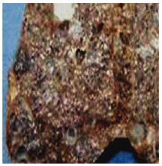
Figure 2 Multiloculated primary splenic cyst.
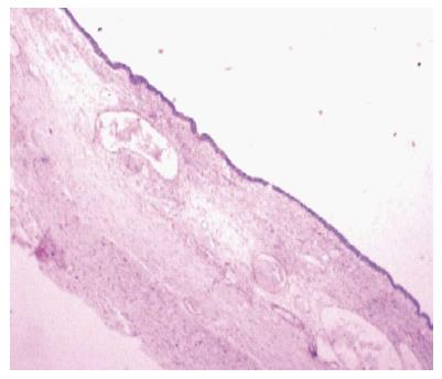
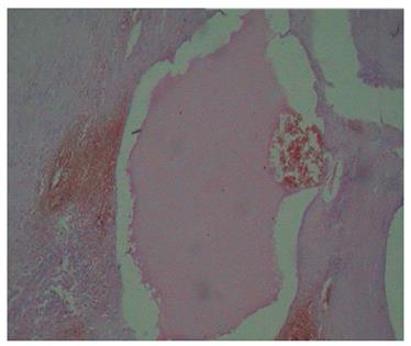
Figure 3 Epidermoid cyst of the spleen (Hematoxylin and eosin staining, × 10).
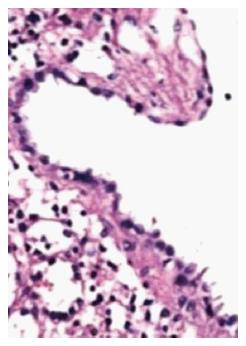
Figure 4 Primary splenic cyst lined with cuboidal to flattened epithelium (Hematoxylin and eosin staining, × 10).
Figure 5 Diffuse lymphangiomatosis of the spleen (Hematoxylin and eosin staining, × 10).
- Citation: Ingle SB, Hinge (Ingle) CR, Patrike S. Epithelial cysts of the spleen: A minireview. World J Gastroenterol 2014; 20(38): 13899-13903
- URL: https://www.wjgnet.com/1007-9327/full/v20/i38/13899.htm
- DOI: https://dx.doi.org/10.3748/wjg.v20.i38.13899


