Copyright
©2014 Baishideng Publishing Group Inc.
World J Gastroenterol. Oct 14, 2014; 20(38): 13879-13892
Published online Oct 14, 2014. doi: 10.3748/wjg.v20.i38.13879
Published online Oct 14, 2014. doi: 10.3748/wjg.v20.i38.13879
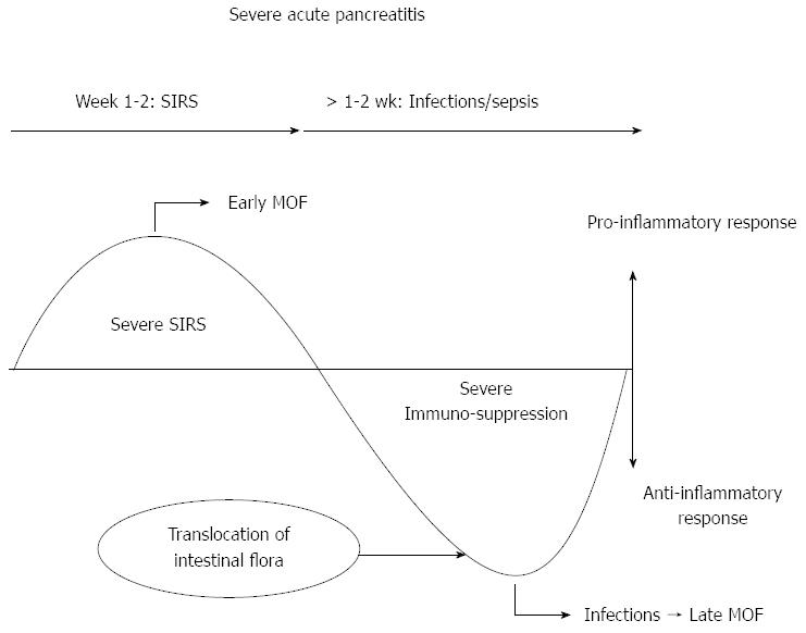
Figure 1 Natural clinical course of severe acute pancreatitis.
SIRS: Systemic inflammatory response syndrome; MOF: Multisystem organ failure.
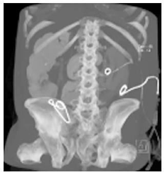
Figure 2 Three catheters inserted percutaneously into the abscess collections formed during the clinical course of necrotizing pancreatitis.
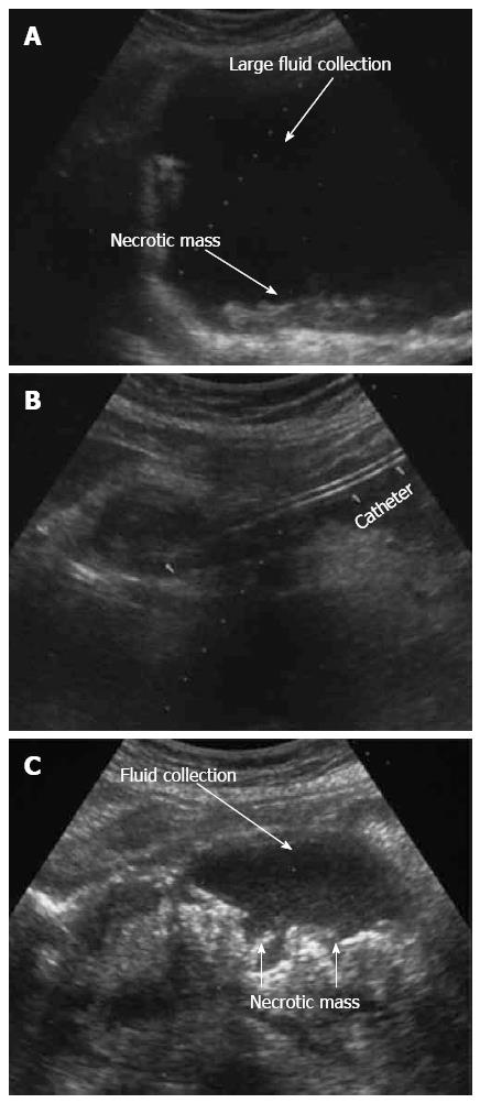
Figure 3 Ultrasound appearance of pancreatic necroses and a large acute fluid collection before and after drainage.
A: Large fluid collection and pancreatic necroses before drainage; B: Catheter in the peripancreatic fluid collection; C: Massive pancreatic necroses with secondary fluid collection.
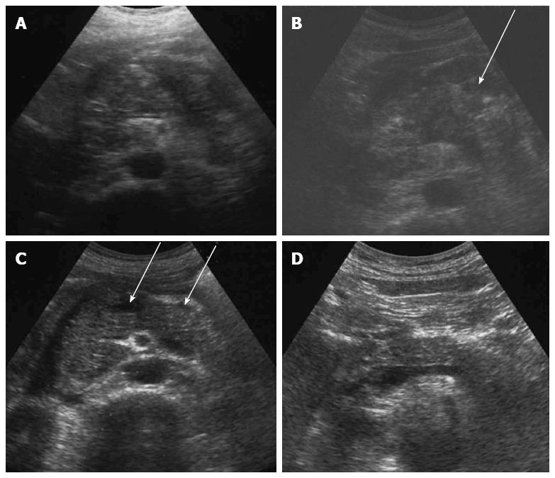
Figure 4 Ultrasound appearance of infected pancreatic necrosis before and after the treatment of acute pancreatitis.
A: Infected pancreatic necrosis (IPN) involved the entire pancreas in the beginning of the disease; B: Liquefied areas in the IPN marked by arrows; C: Small necroses and liquid collections around the pancreas 2 mo after the beginning of treatment marked by arrows; D: Normal appearance of the pancreas 6 mo after the beginning of treatment.
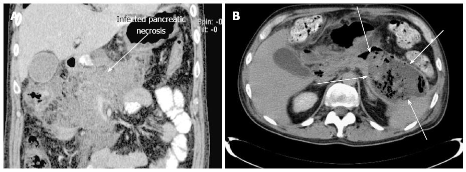
Figure 5 Computed tomography.
A: Computed tomography (CT) appearance of the infected pancreatic necrosis, which involves the entire pancreas (marked by an arrow); B: CT appearance of a large pancreatic walled-off necrosis in the tail of the pancreas (marked by arrows).
- Citation: Zerem E. Treatment of severe acute pancreatitis and its complications. World J Gastroenterol 2014; 20(38): 13879-13892
- URL: https://www.wjgnet.com/1007-9327/full/v20/i38/13879.htm
- DOI: https://dx.doi.org/10.3748/wjg.v20.i38.13879


