©2014 Baishideng Publishing Group Inc.
World J Gastroenterol. Oct 7, 2014; 20(37): 13273-13283
Published online Oct 7, 2014. doi: 10.3748/wjg.v20.i37.13273
Published online Oct 7, 2014. doi: 10.3748/wjg.v20.i37.13273
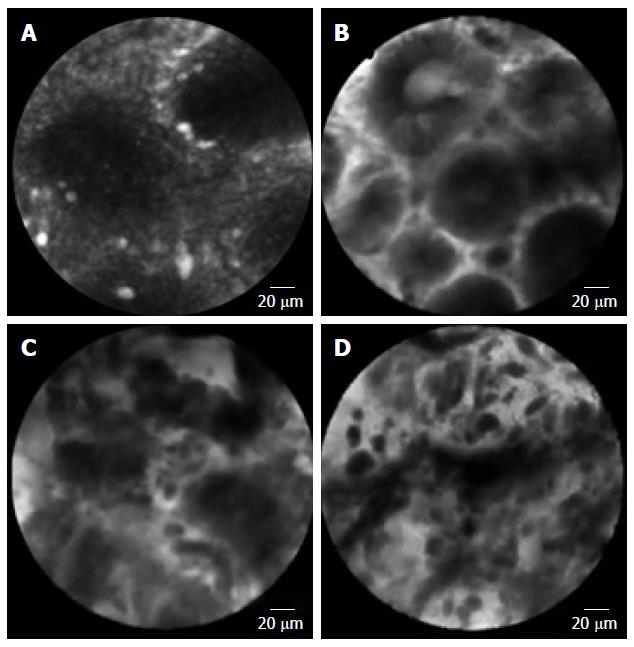
Figure 1 Features of confocal endomicroscopy.
A: Normal gastric epithelium, round pattern of normal crypts is observed; B: Dysplasia, dark epithelium with irregular and varying thickness is observed; C: Differentiated adenocarcinoma, disorganized epithelium with dark and irregular glands is observed; D: Undifferentiated adenocarcinoma, dark and irregular cells with no identifiable glandular structures are observed.
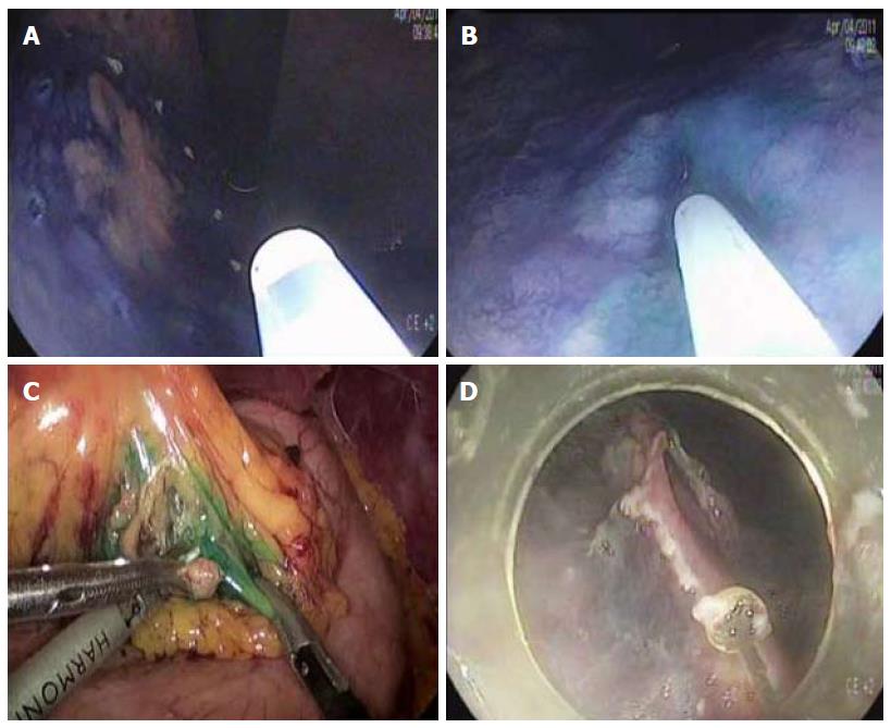
Figure 2 Endoscopic submucosal dissection with sentinel node navigation.
A: Marking for endoscopic submucosal dissection is performed around the tumor; B: Indocyanine green is injected into the submucosal layer around the tumor for sentinel node navigation; C: Sentinel node harvest is performed by laparoscopic pick-up biopsy; D: Endoscopic submucosal dissection is performed.
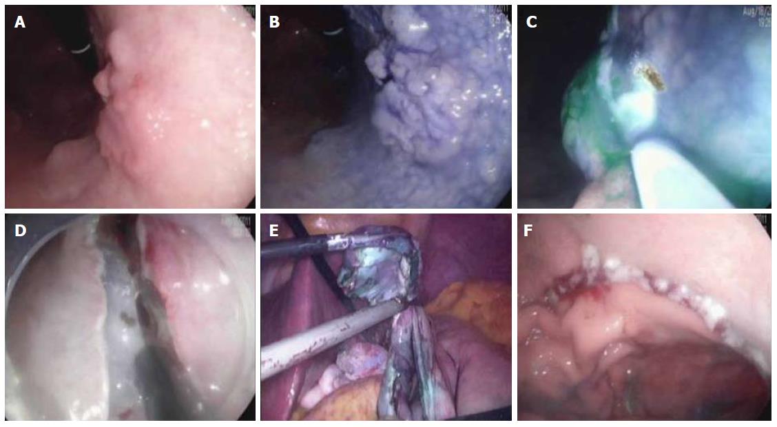
Figure 3 Full-thickness gastric resection.
A: An elevated lesion is noted at the lesser curvature of upper body; B: The lesion becomes distinct by chromoendoscopy using acetic acid and indigocarmin; C: For sentinel node navigation, indocyanine green is injected into the submucosal layer after marking around the tumor; D: Endoscopic full-thickness resection is performed after sentinel node harvest and regional lymph node dissection; E: Final resection is performed with laparoscopy; F: Gastric closure is achieved with laparoscopy.
- Citation: Kim MY, Cho JH, Cho JY. Ever-changing endoscopic treatment for early gastric cancer: Yesterday-today-tomorrow. World J Gastroenterol 2014; 20(37): 13273-13283
- URL: https://www.wjgnet.com/1007-9327/full/v20/i37/13273.htm
- DOI: https://dx.doi.org/10.3748/wjg.v20.i37.13273


