©2014 Baishideng Publishing Group Inc.
World J Gastroenterol. Sep 21, 2014; 20(35): 12668-12672
Published online Sep 21, 2014. doi: 10.3748/wjg.v20.i35.12668
Published online Sep 21, 2014. doi: 10.3748/wjg.v20.i35.12668
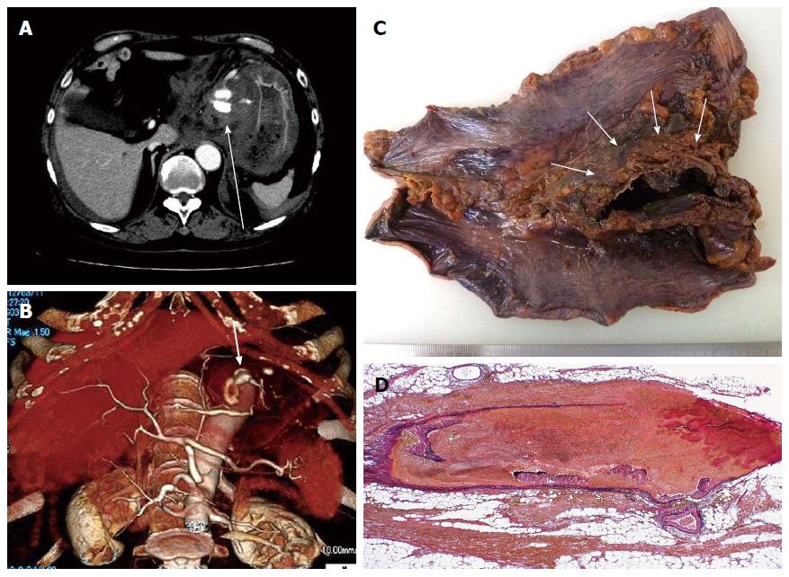
Figure 1 Radiological and pathological findings of the gastric artery aneurysm.
A: Abdominal computed tomography (CT) showing a hematoma in the lesser omentum (arrow); B: CT angiography showing the aneurysm of the left gastric artery (arrow); C: Surgical specimen of the stomach. In addition to the hematoma in the lesser omentum (arrows), marked subserosal hemorrhage is seen; D: Histological findings of the aneurysm. The primary pathologic change is dissection and fragmentation of the arterial wall. No inflammatory change is seen (Elastica van Gieson stain; original magnification, × 20).
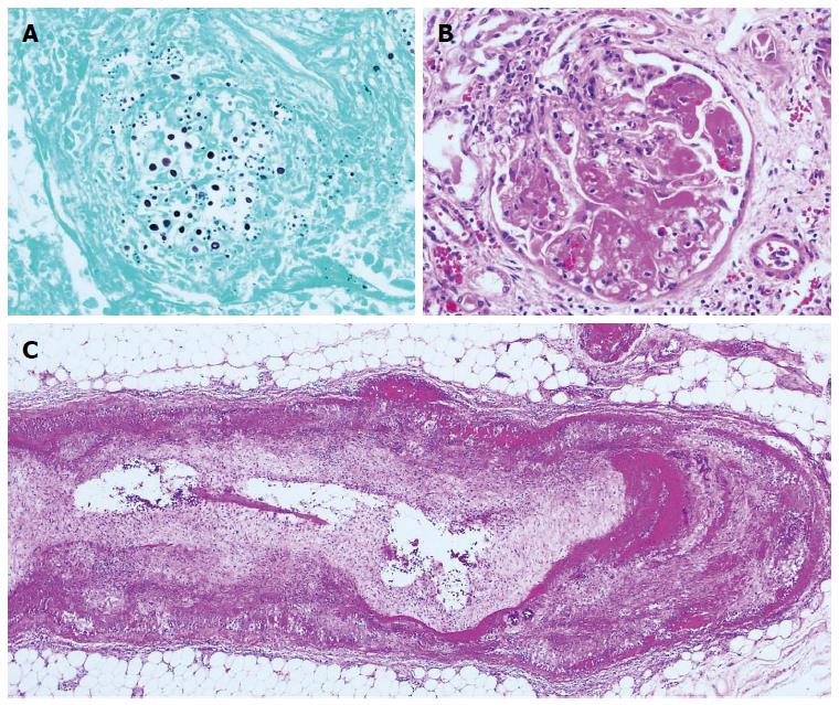
Figure 2 Representative histological findings in the autopsy samples.
A: Cryptococcus in the lung (Grocott stain, original magnification, × 400); B: Necrotizing glomerulitis (Hematoxylin-eosin, original magnification, × 400); C: Necrotizing arteritis of the urinary bladder (Hematoxylin-eosin, original magnification, × 40).
- Citation: Ikura Y, Kadota T, Watanabe S, Arimoto A, Nishioka E. Ruptured gastric artery aneurysm: An uncommon manifestation of microscopic polyangiitis. World J Gastroenterol 2014; 20(35): 12668-12672
- URL: https://www.wjgnet.com/1007-9327/full/v20/i35/12668.htm
- DOI: https://dx.doi.org/10.3748/wjg.v20.i35.12668


