©2014 Baishideng Publishing Group Inc.
World J Gastroenterol. Sep 21, 2014; 20(35): 12657-12661
Published online Sep 21, 2014. doi: 10.3748/wjg.v20.i35.12657
Published online Sep 21, 2014. doi: 10.3748/wjg.v20.i35.12657
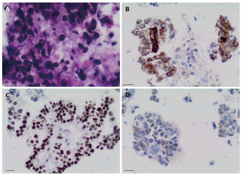
Figure 1 Fine needle aspiration biopsy of enlarged porta hepatis node.
A: Papanicolaou staining showed multiple intermediate-sized dysplastic cells with vacuolated cytoplasm and occasional markedly-enlarged malignant nuclei-forming lumina (× 40 magnification); B: Immunohistochemistry showed that tumor cells stained positive for alpha-fetoprotein (× 20 magnification); C: Tumor cells showed strong nuclear immunostaining for caudal-related homeobox transcription factor 2 (× 20 magnification); D: Staining for hepatocyte-specific antigen was unequivocal (× 20 magnification).
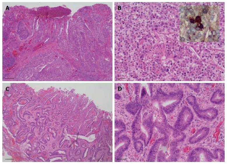
Figure 2 Colectomy specimen showed two synchronous tumors with divergent histologic differentiation.
A: The larger tumor was poorly differentiated and composed of sheets of polygonal-shaped cells with granular eosinophilic cytoplasm, centrally located nuclei and prominent nucleoli (hematoxylin and eosin staining, × 40 magnification); B: Larger tumor at × 200 magnification, inset shows immunostaining for alpha-fetoprotein; C: The second tumor demonstrated a more typical colonic morphologic differentiation, characterized by infiltrating submucosal glands (hematoxylin and eosin staining, × 40 magnification); D: Second tumor at × 200 magnification.
- Citation: Chen Y, Schaeffer DF, Yoshida EM. Hepatoid adenocarcinoma of the colon in a patient with inflammatory bowel disease. World J Gastroenterol 2014; 20(35): 12657-12661
- URL: https://www.wjgnet.com/1007-9327/full/v20/i35/12657.htm
- DOI: https://dx.doi.org/10.3748/wjg.v20.i35.12657


