©2014 Baishideng Publishing Group Inc.
World J Gastroenterol. Sep 21, 2014; 20(35): 12621-12627
Published online Sep 21, 2014. doi: 10.3748/wjg.v20.i35.12621
Published online Sep 21, 2014. doi: 10.3748/wjg.v20.i35.12621
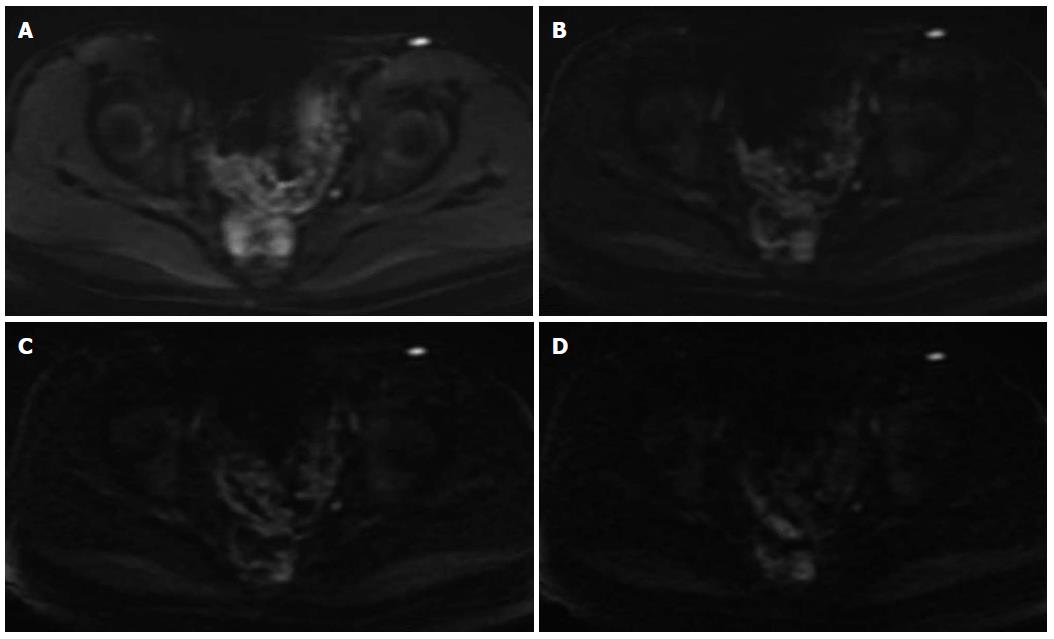
Figure 2 Images obtained in a 34-year-old man.
There was high signal intensity in the sigmoid colon and rectum when b value 800 s/mm2 (A) was chosen. But when b value 1500 s/mm2 to 2500 s/mm2 (B-D) was chosen, the high signal was apparently depressed. It was confirmed that the sigmoid colon and rectum were normal by colonic endoscopy.
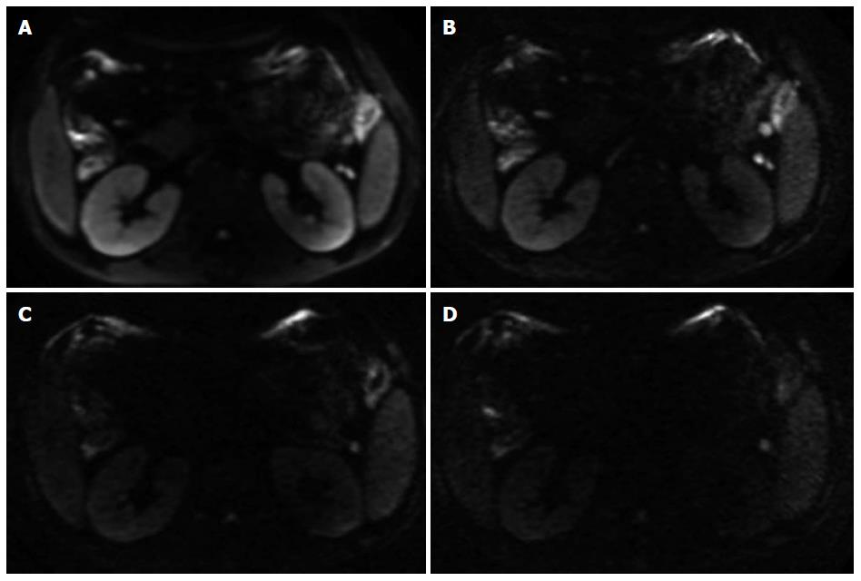
Figure 1 Diffusion-weighted imaging images obtained in a 28-year-old man.
The whole colonic inflammation was identified by endoscopy. The high signal intensity of colonic liver flexure and splenic flexure could be recognized on b = 800 s/mm2 (A) and b = 1500 s/mm2 images (B). But when b = 2000 s/mm2 or b = 2500 s/mm2 (C and D) was chosen, the signal intensity was depressed and these two segments were misdiagnosed as negative.
-
Citation: Feng Q, Yan YQ, Zhu J, Tong JL, Xu JR. Optimal
b value of diffusion-weighted imaging on a 3.0T magnetic resonance scanner in Crohn’s disease. World J Gastroenterol 2014; 20(35): 12621-12627 - URL: https://www.wjgnet.com/1007-9327/full/v20/i35/12621.htm
- DOI: https://dx.doi.org/10.3748/wjg.v20.i35.12621


