Copyright
©2014 Baishideng Publishing Group Inc.
World J Gastroenterol. Aug 7, 2014; 20(29): 9998-10007
Published online Aug 7, 2014. doi: 10.3748/wjg.v20.i29.9998
Published online Aug 7, 2014. doi: 10.3748/wjg.v20.i29.9998
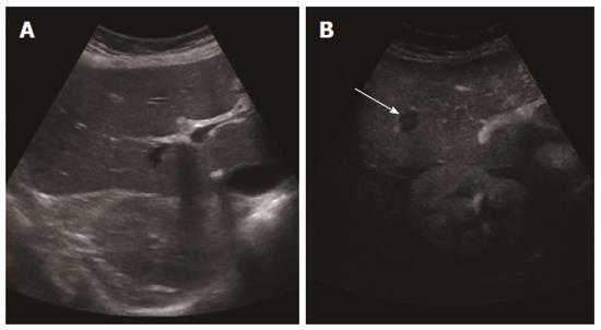
Figure 1 Baseline ultrasound did not detect any lesion (A), while contrast enhanced ultrasound clearly showed one hypoenhancing lesion (B) (arrow).
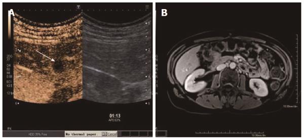
Figure 2 Both contrast enhanced ultrasound (A), (arrow showing the metastases) and BOPTA-enhanced magnetic resonance imaging detected a liver metastasis (B).
- Citation: Cantisani V, Grazhdani H, Fioravanti C, Rosignuolo M, Calliada F, Messineo D, Bernieri MG, Redler A, Catalano C, D’Ambrosio F. Liver metastases: Contrast-enhanced ultrasound compared with computed tomography and magnetic resonance. World J Gastroenterol 2014; 20(29): 9998-10007
- URL: https://www.wjgnet.com/1007-9327/full/v20/i29/9998.htm
- DOI: https://dx.doi.org/10.3748/wjg.v20.i29.9998


