©2014 Baishideng Publishing Group Inc.
World J Gastroenterol. Jul 28, 2014; 20(28): 9549-9555
Published online Jul 28, 2014. doi: 10.3748/wjg.v20.i28.9549
Published online Jul 28, 2014. doi: 10.3748/wjg.v20.i28.9549
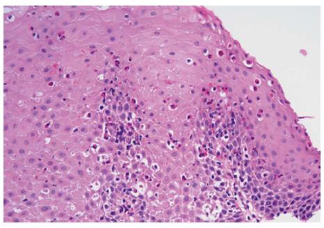
Figure 1 Reactive squamous mucosa with marked increased intraepithelial eosinophilia involving the entire thickness of the epithelium, occasional eosinophilic microabscesses and degranulation of eosinophils (HE stain, × 400).
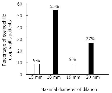
Figure 2 Maximal size of dilation used in 22 eosinophilic esophagitis patients.
- Citation: Ukleja A, Shiroky J, Agarwal A, Allende D. Esophageal dilations in eosinophilic esophagitis: A single center experience. World J Gastroenterol 2014; 20(28): 9549-9555
- URL: https://www.wjgnet.com/1007-9327/full/v20/i28/9549.htm
- DOI: https://dx.doi.org/10.3748/wjg.v20.i28.9549


