©2014 Baishideng Publishing Group Inc.
World J Gastroenterol. Jul 14, 2014; 20(26): 8745-8750
Published online Jul 14, 2014. doi: 10.3748/wjg.v20.i26.8745
Published online Jul 14, 2014. doi: 10.3748/wjg.v20.i26.8745
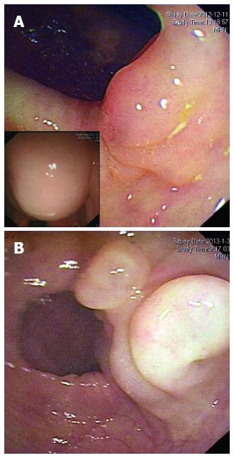
Figure 1 Endoscopic findings for the two cases.
A round, semi-transparent and wide-based submucosal lesion at the ascending colon in Case 1 (A) and a sharply marginated, lobular submucosal lesion at the sigmoid colon in Case 2 (B) with gentle slopes and smooth surfaces are noted. Left bottom of Figure 1A shows that when the patient's position was altered, the shape of the mass changed and was fluctuant on palpation.
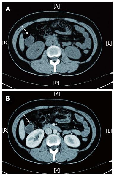
Figure 2 Abdominopelvic computed tomography scan for Case 1 revealed a cystic mass (indicated by white arrow) located at the ascending colon, which was visible in a non-contrast scan (A) and was not enhanced after the arterial phase (B).
A: Anterior; R: Right; L: Left; P: Posterior.
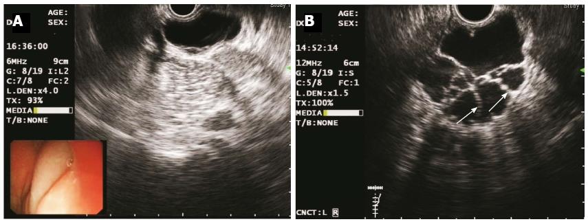
Figure 3 Endoscopic ultrasonography images of the two cases obtained using a catheter EUS probe.
A: Elevated lesions as echo-free, homogenous cysts in Case 1 (frequency 6 MHz); B: Multiple septal walls (indicated by white arrows) in Case 2 (frequency 12 MHz); EUS revealed that both lesions were located in the third (submucosal) layer.
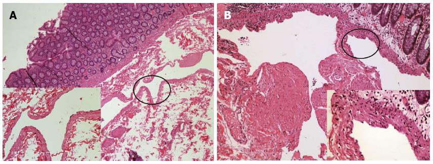
Figure 4 Histopathological examination of the two cases after hematoxylin and eosin stain (at × 40 magnification for low power field images).
A: Case 1 revealed a dilated submucosal cyst lined with a single layer of endothelial cells [left bottom, hematoxylin and eosin (HE) stain, × 200]; B: Case 2 displayed lymphatic vessels of varied sizes, which were covered by a flattened layer of lymphatic epithelium. The smooth muscle layer of the vessels is also noted (right bottom, HE stain, × 400).
- Citation: Zhuo CH, Shi DB, Ying MG, Cheng YF, Wang YW, Zhang WM, Cai SJ, Li XX. Laparoscopic segmental colectomy for colonic lymphangiomas: A definitive, minimally invasive surgical option. World J Gastroenterol 2014; 20(26): 8745-8750
- URL: https://www.wjgnet.com/1007-9327/full/v20/i26/8745.htm
- DOI: https://dx.doi.org/10.3748/wjg.v20.i26.8745


