Copyright
©2014 Baishideng Publishing Group Inc.
World J Gastroenterol. Jul 14, 2014; 20(26): 8729-8735
Published online Jul 14, 2014. doi: 10.3748/wjg.v20.i26.8729
Published online Jul 14, 2014. doi: 10.3748/wjg.v20.i26.8729
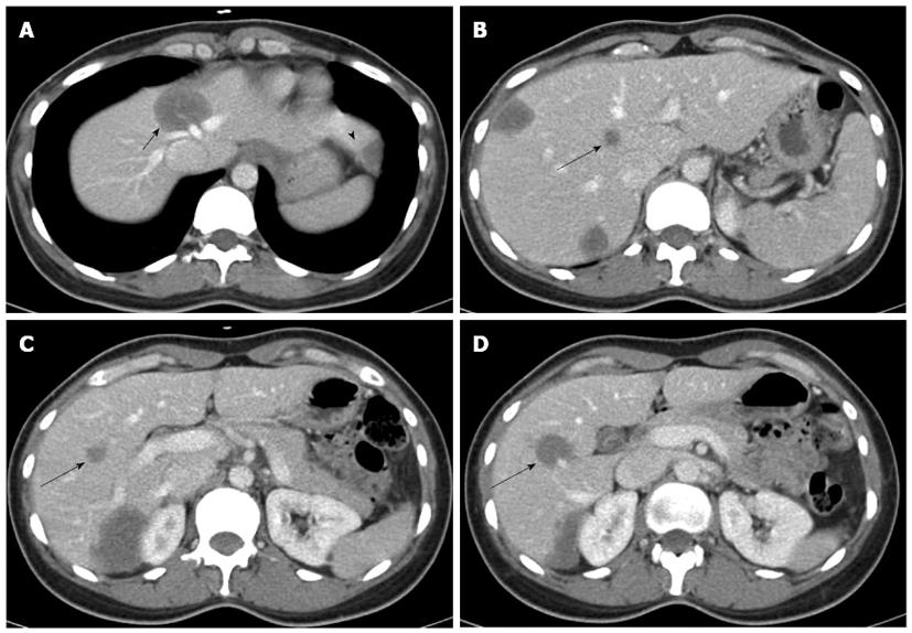
Figure 1 Abdominal computed tomography revealed a total of 7 lesions located in both lobes.
A: The tumor of segment 4 (short arrow) hinders the possibility of right hemihepatectomy, because it invades the roots of the middle and left hepatic veins (arrowhead indicates the tumor of segment 3); B-D: The 3 deep-seated tumors in the right lobe (long arrows) hinder the possibility of left hemihepatectomy.
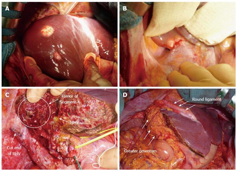
Figure 2 Intraoperative images.
A: The tumors were well-circumscribed, whitish, and firm; B: The tumor of segment 6 invaded Gerota’s fascia; C: After right lobectomy, the tumor of segment 4 was very close to the cut surface of the liver; D: A flap of the greater omentum (arrows) was sutured over the cut surface of the liver to cover the tumor of segment 4. MHV: Middle hepatic vein; RHV: Right hepatic vein.
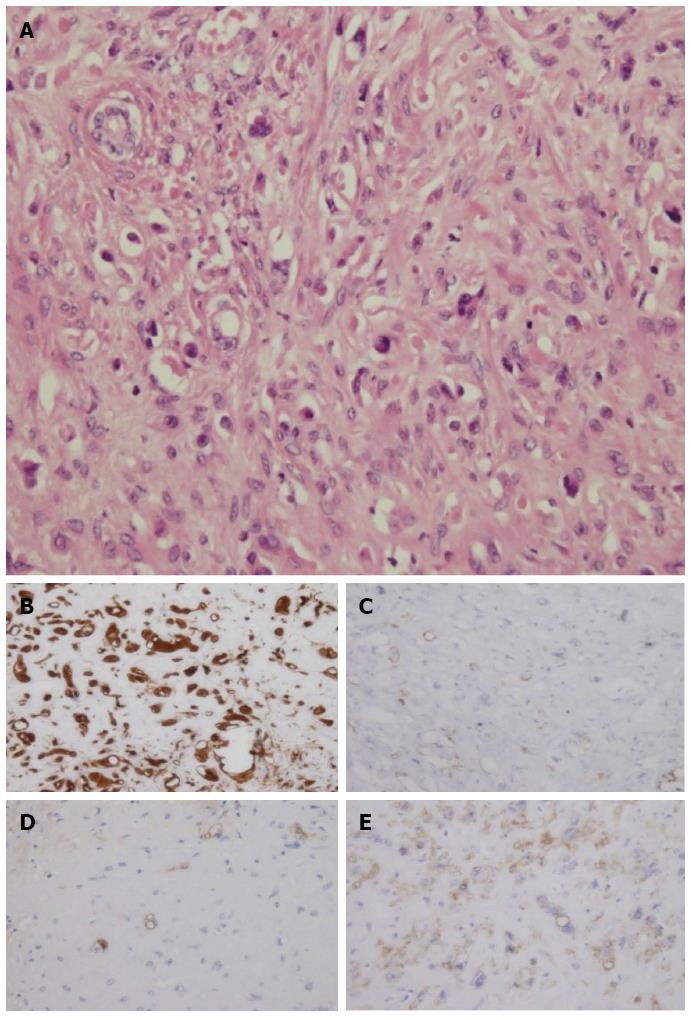
Figure 3 Pathological findings.
A: The specimen was composed of many atypical vacuolatd and dendritic cells; B-E: The tumor cells were immunoreactive for vimentin (B), factor VIII-related antigen (C), CD31 (D), and CD34 (E).
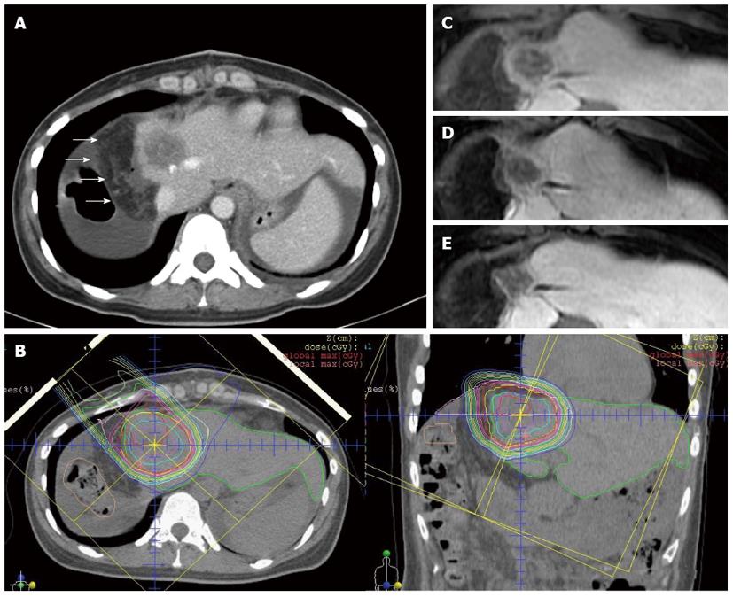
Figure 4 Treatment course.
A: Post-operative abdominal computed tomography (CT) showed the greater omental flap (arrows) around the tumor, maintaining a sufficient space between the tumor and the gastrointestinal tract; B: Treatment plan for carbon-ion radiotherapy with a total dose of 76 GyE in 20 fractions. Isodose lines demonstrate 100% of the prescribed dose at the center and decreasing by 10% of the dose from the inside to the outside. Owing to the greater omental flap, the tumor was entirely irradiated with full dose, and the gastrointestinal tract was successfully spared; C: Comparison of abdominal CT and magnetic resonance imaging scans taken over a period of 24 mo after carbon-ion radiotherapy indicated shrinkage of the tumor with decreasing tumor vascularity. The maximum tumor diameter was 35 mm before treatment, and tumor size decreased thereafter to 27 mm after 6 mo; D: Tumor size of 26 mm after 12 mo; E: Tumor size of 24 mm after 24 mo.
- Citation: Komatsu S, Iwasaki T, Demizu Y, Terashima K, Fujii O, Takebe A, Toyokawa A, Teramura K, Fukumoto T, Ku Y, Fuwa N. Two-stage treatment with hepatectomy and carbon-ion radiotherapy for multiple hepatic epithelioid hemangioendotheliomas. World J Gastroenterol 2014; 20(26): 8729-8735
- URL: https://www.wjgnet.com/1007-9327/full/v20/i26/8729.htm
- DOI: https://dx.doi.org/10.3748/wjg.v20.i26.8729


