©2014 Baishideng Publishing Group Inc.
World J Gastroenterol. Jul 14, 2014; 20(26): 8717-8721
Published online Jul 14, 2014. doi: 10.3748/wjg.v20.i26.8717
Published online Jul 14, 2014. doi: 10.3748/wjg.v20.i26.8717
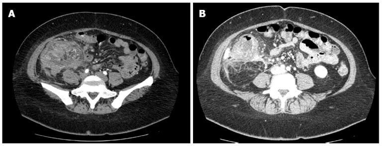
Figure 1 Contrast enhanced computed tomography showing a large mass in the right abdominal quadrant (A), and inflammatory involvement of retroperitoneal fat and local lymph nodes (B).
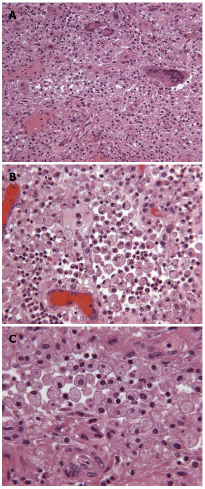
Figure 2 Pathological findings.
Hematoxylin and eosin staining of the intestinal wall of the ileocecal valve and appendix showing heavy infiltration of a large amount of lipid-laden macrophages in which scattered multinucleated giant cells are shown (A, × 160). In the field, numerous ectatic small blood vessels consistent with the edematous features of the wall are visible (B, × 250; C, × 400).
- Citation: Chieco PA, Antolino L, Giaccaglia V, Centanini F, Cunsolo GV, Sparagna A, Uccini S, Ziparo V. Acute abdomen: Rare and unusual presentation of right colic xanthogranulomatosis. World J Gastroenterol 2014; 20(26): 8717-8721
- URL: https://www.wjgnet.com/1007-9327/full/v20/i26/8717.htm
- DOI: https://dx.doi.org/10.3748/wjg.v20.i26.8717


