©2014 Baishideng Publishing Group Inc.
World J Gastroenterol. Jul 14, 2014; 20(26): 8458-8470
Published online Jul 14, 2014. doi: 10.3748/wjg.v20.i26.8458
Published online Jul 14, 2014. doi: 10.3748/wjg.v20.i26.8458
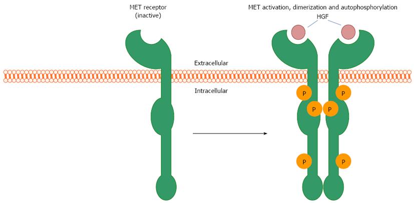
Figure 1 The mesenchymal-epithelial transition factor receptor functions as a transmembrane tyrosine kinase receptor.
Ligand binding from hepatocyte growth factor (HGF)/scatter factor induces receptor dimerization and autophosphorylation of intracellular tyrosine residues, which serves as a catalytic site for the SH2 domains of numerous cytosolic signaling proteins. MET: Mesenchymal-epithelial transition factor.
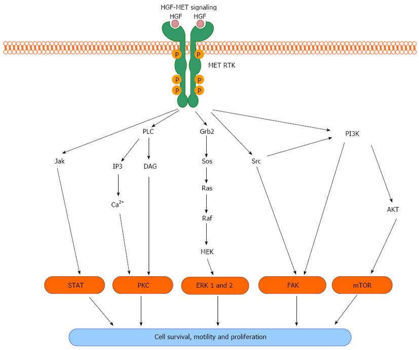
Figure 2 Hepatocyte growth factor activation of the mesenchymal-epithelial transition tyrosine kinase receptor induces a pleiotropic response involving a host of intracellular signaling to induce cell survival, migration and proliferation.
HGF: Hepatocyte growth factor; MET: Mesenchymal-epithelial transition factor; RTK: Receptor tyrosine kinase; JAK: Janus kinase; STAT: Signal transducer and activator of transcription; PLC: Phospholipase C; IP3: Inositol triphosphate; DAG: Diacylglycerol; Ca2+: Calcium; PKC: Protein kinase C; Grb2: Growth factor receptor-bound protein 2; Sos: Son of sevenless homolog; Ras: Harvey rat sarcoma viral oncogene homolog; Raf: Rapidly accelerating fibrosarcoma; MEK: Mitogen activated protein kinase kinase; ERK: Extracellular-signal-regulated kinase; FAK: Focal adhesion kinase; PI3K: Phosphoinositide 3-kinase; AKT: Protein kinase B; mTOR: Mammalian target of rapamycin.
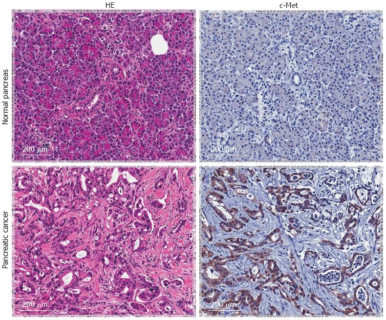
Figure 3 Immunoperoxidase staining.
Immunoperoxidase staining of formalin fixed, paraffin embedded human pancreatic specimens demonstrate over expression of c-Met receptor in pancreatic cancer patients when compared to adjacent normal pancreatic tissue controls (right panel). HE staining demonstrate histological confirmation of diseased (pancreatic cancer) or normal tissue (left panel).
- Citation: Delitto D, Vertes-George E, Hughes SJ, Behrns KE, Trevino JG. c-Met signaling in the development of tumorigenesis and chemoresistance: Potential applications in pancreatic cancer. World J Gastroenterol 2014; 20(26): 8458-8470
- URL: https://www.wjgnet.com/1007-9327/full/v20/i26/8458.htm
- DOI: https://dx.doi.org/10.3748/wjg.v20.i26.8458


