©2014 Baishideng Publishing Group Inc.
World J Gastroenterol. Jun 7, 2014; 20(21): 6534-6540
Published online Jun 7, 2014. doi: 10.3748/wjg.v20.i21.6534
Published online Jun 7, 2014. doi: 10.3748/wjg.v20.i21.6534
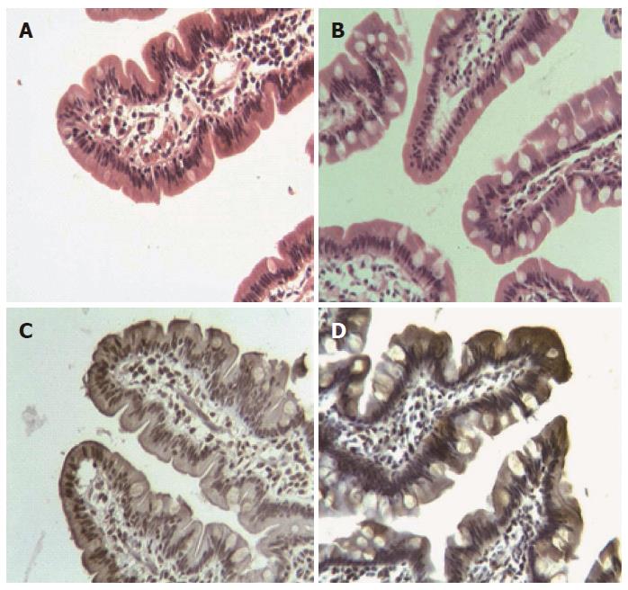
Figure 1 Morphological and immunohistochemical changes in the jejunal villus of patients subjected to Roux-en-Y gastric bypass.
A and B are jejunal villus stained with haematoxylin-eosin at 0 and 6 mo after surgery, respectively. Note the preserved normal morphological pattern together with an increase of goblet cells. C and D show immunohistochemical staining for divalent metal transporter 1 during the same period, with an evident increase of such staining in the cytoplasm of epithelial cells at the apex of the villi.
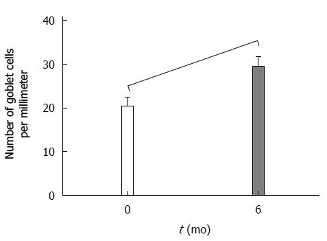
Figure 2 Change in the number of goblet cells in the proximal jejunum 6 mo after Roux-en-Y gastric bypass.
Wilcoxon test: Z = -2.47; P = 0.013.
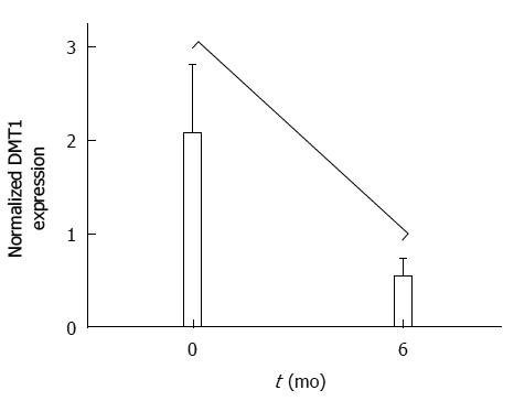
Figure 3 Change in expression of divalent metal transporter 1 in the proximal jejunum 6 mo after Roux-en-Y gastric bypass.
Wilcoxon test: Z = 2.04; P = 0.04.
- Citation: Marambio A, Watkins G, Castro F, Riffo A, Zúñiga R, Jans J, Villanueva ME, Díaz G. Changes in iron transporter divalent metal transporter 1 in proximal jejunum after gastric bypass. World J Gastroenterol 2014; 20(21): 6534-6540
- URL: https://www.wjgnet.com/1007-9327/full/v20/i21/6534.htm
- DOI: https://dx.doi.org/10.3748/wjg.v20.i21.6534


