Copyright
©2014 Baishideng Publishing Group Co.
World J Gastroenterol. Apr 28, 2014; 20(16): 4835-4838
Published online Apr 28, 2014. doi: 10.3748/wjg.v20.i16.4835
Published online Apr 28, 2014. doi: 10.3748/wjg.v20.i16.4835
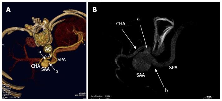
Figure 1 Three-dimensional computed tomography reconstruction.
A: A 3-cm splenic artery aneurysm in the proximal splenic artery with afferent (a) and efferent (b) artery; B: Angiography demonstrating a 3 cm splenic artery aneurysm at the same location with afferent (a) and efferent (b) artery. AO: Abdominal aorta; CA: Celiac artery; CHA: Common hepatic artery; SAA: Splenic artery aneurysm; SPA: Splenic artery.
Figure 2 Intraoperative imaging.
A: The position of the splenic artery aneurysm with its afferent (a) and efferent (b) artery; B: Both afferent (a) and efferent (b) arteries of the aneurysm were occluded by the ligaclips; C: Reverse side of the splenic artery aneurysm showing no collateral vessels connecting with the aneurysm. CHA: Common hepatic artery; SAA: Splenic artery aneurysm; SPA: Splenic artery. LGA: Left gastric artery; PV: Portal vein; SPV: Splenic vein.
Figure 3 Postoperative imaging.
A: Ultrasound showing a 3 cm hypoechoic mass between the proximal splenic artery and the celiac trunk; B: Ultrasound showing no color flow signals in the hypoechoic mass; C: Three-dimensional CT reconstruction revealing the proximal splenic artery without aneurysm recurrence and without collateral vessels supplying the spleen (c). AO: Abdominal aorta; CA: Celiac artery; CHA: Common hepatic artery; LGA: Left gastric artery.
- Citation: Wei YH, Xu JW, Shen HP, Zhang GL, Ajoodhea H, Zhang RC, Mou YP. Laparoscopic ligation of proximal splenic artery aneurysm with splenic function preservation. World J Gastroenterol 2014; 20(16): 4835-4838
- URL: https://www.wjgnet.com/1007-9327/full/v20/i16/4835.htm
- DOI: https://dx.doi.org/10.3748/wjg.v20.i16.4835


