Copyright
©2014 Baishideng Publishing Group Co.
World J Gastroenterol. Apr 28, 2014; 20(16): 4597-4606
Published online Apr 28, 2014. doi: 10.3748/wjg.v20.i16.4597
Published online Apr 28, 2014. doi: 10.3748/wjg.v20.i16.4597
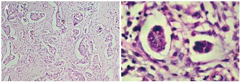
Figure 1 Characterization of typical invasive micropapillary carcinoma structures.
A: Invasive micropapillary carcinoma in the invasive edges of the tumor; B: Morphologically, clusters of small rounded neoplastic cells without fibrous-vascular core. Hematoxylin and eosin stain, × 40, × 400, respectively.
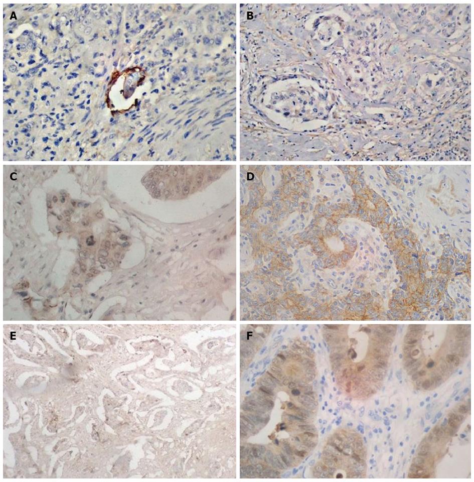
Figure 2 Immunohistochemical characteristics of invasive micropapillary carcinoma structures.
Positive immunoreaction of lymphatic endothelial cell performed by D2-40 (A) and lack of reaction in invasive micropapillary carcinoma (IMPC) structures (B). Immunohistochemical expression of E-catherin in membrane of conventional adenocarcinoma (C) and in cytoplasm of IMPC (D). The cytoplasmic expression of Beta-catenin was observed more frequent in typical adenocarcinoma (F) compare to IMPC (E). Magnification × 400.
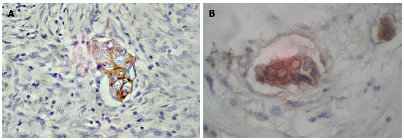
Figure 3 Immunohistochemical characteristics of invasive micropapillary carcinoma of colon.
Positive reaction of CK20 (A) and CEA (B). Magnification × 400.
- Citation: Guzińska-Ustymowicz K, Niewiarowska K, Pryczynicz A. Invasive micropapillary carcinoma: A distinct type of adenocarcinomas in the gastrointestinal tract. World J Gastroenterol 2014; 20(16): 4597-4606
- URL: https://www.wjgnet.com/1007-9327/full/v20/i16/4597.htm
- DOI: https://dx.doi.org/10.3748/wjg.v20.i16.4597


