©2014 Baishideng Publishing Group Co.
World J Gastroenterol. Apr 7, 2014; 20(13): 3628-3634
Published online Apr 7, 2014. doi: 10.3748/wjg.v20.i13.3628
Published online Apr 7, 2014. doi: 10.3748/wjg.v20.i13.3628
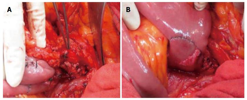
Figure 1 An intraoperative photograph of the mesenteric vessels (A), and of anastomosis on the left side of the mesenteric vessels (B).
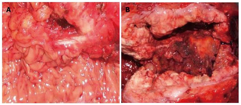
Figure 2 A well-encapsulated gastrointestinal stromal tumor with a normal mucous membrane(A), and an ulcerative carcinoma that has infiltrated into the adjacent tissue (B).
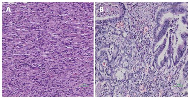
Figure 3 Gastrointestinal stromal tumors with spindle cell differentiation (A) (× 20), and carcinoma with moderate-to-poor differentiation (B) (× 20).
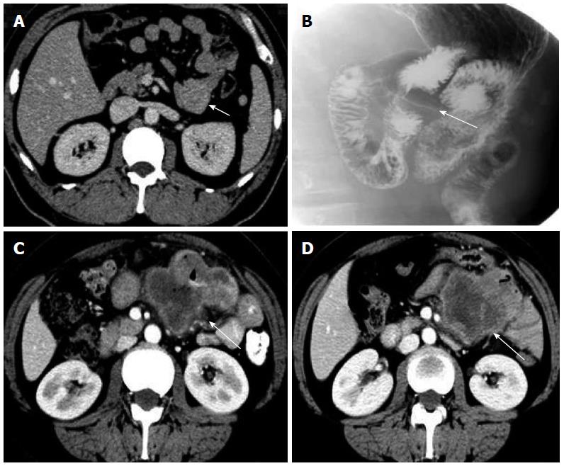
Figure 4 Computed tomography scan.
A, B: Computed tomography (CT) scan and upper gastrointestinal radiography for gastrointestinal stromal tumors of the angle of Treitz (arrow); C, D: CT scan of adenocarcinoma of the angle of Treitz before (C) and after (D) chemotherapy (arrow).
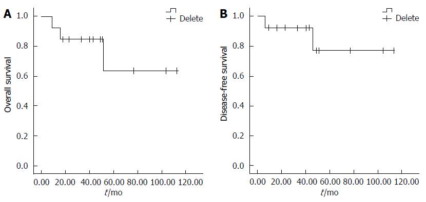
Figure 5 Kaplan-Meier analysis demonstrates the overall (A) and disease-free survival (B) rates for the 13 patients with tumors of the angle of Treitz.
- Citation: Xie YB, Liu H, Cui L, Xing GS, Yang L, Sun YM, Bai XF, Zhao DB, Wang CF, Tian YT. Tumors of the angle of Treitz: A single-center experience. World J Gastroenterol 2014; 20(13): 3628-3634
- URL: https://www.wjgnet.com/1007-9327/full/v20/i13/3628.htm
- DOI: https://dx.doi.org/10.3748/wjg.v20.i13.3628


