©2014 Baishideng Publishing Group Co.
World J Gastroenterol. Mar 14, 2014; 20(10): 2641-2652
Published online Mar 14, 2014. doi: 10.3748/wjg.v20.i10.2641
Published online Mar 14, 2014. doi: 10.3748/wjg.v20.i10.2641
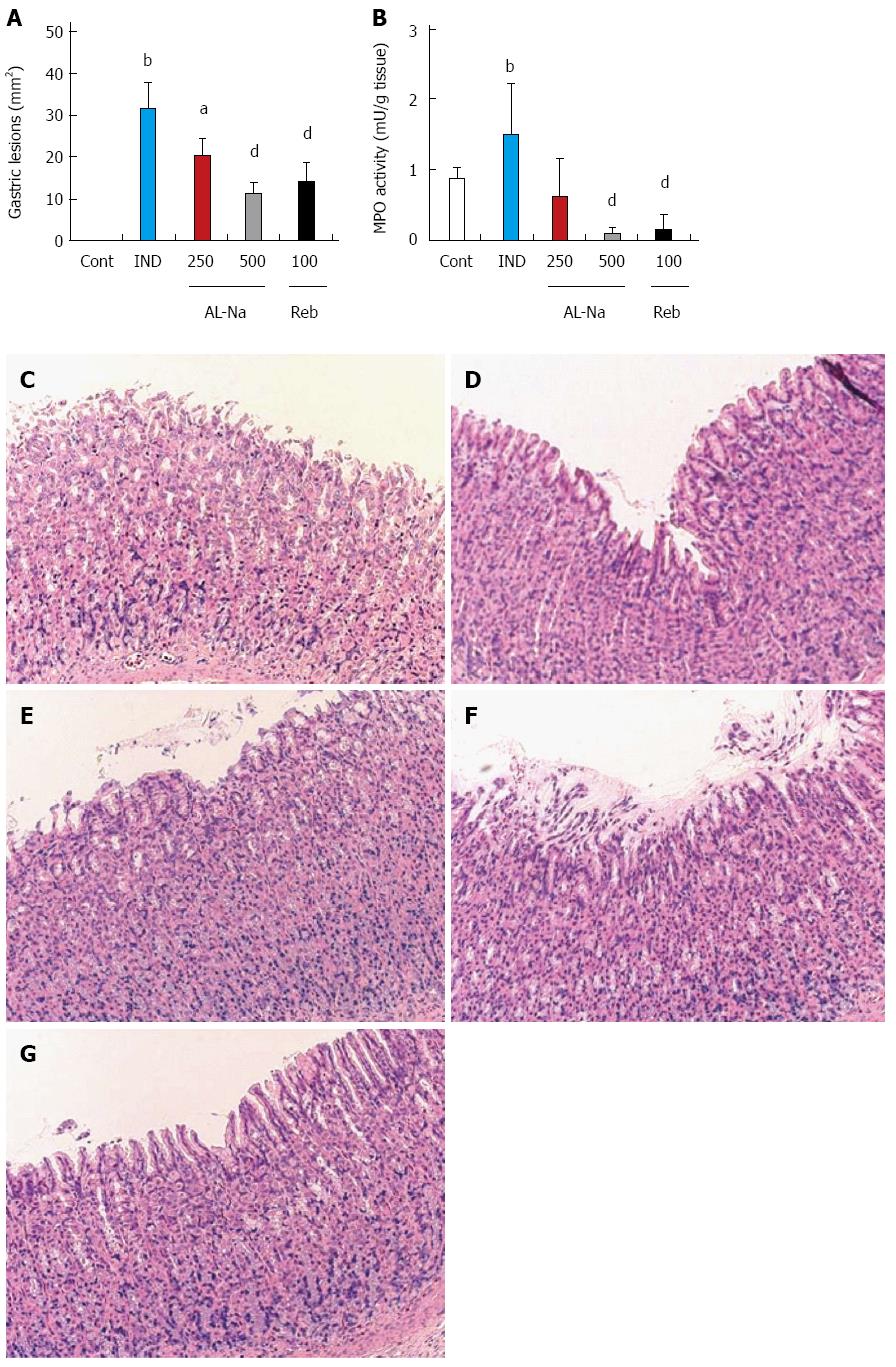
Figure 1 Effects of drugs on indomethacin-induced gastric lesions.
Animals were given indomethacin (25 mg/kg, po) and killed 6 h later. AL-Na (250 and 500 mg/kg) or Reb (100 mg/kg) was given orally at 30 min before administration of indomethacin. The lesion areas (A) and myeloperoxidase (MPO) activity were measured (B). C: Hematoxylin and eosin stained microscopic observations of the rat gastric mucosa of the control (Cont) group; D: Indomethacin (IND); E: AL-Na (250 mg/kg); F: AL-Na (500 mg/kg); G: Reb (100 mg/kg). Each column and vertical bar represents the mean ± SE (n = 8). Significantly different from the Cont group at bP < 0.01 (Student’s t-test). Significantly different from the IND group at aP < 0.05 and dP < 0.01, respectively (Dunnett’s test). AL-Na: Sodium alginate; Reb: Rebamipide.
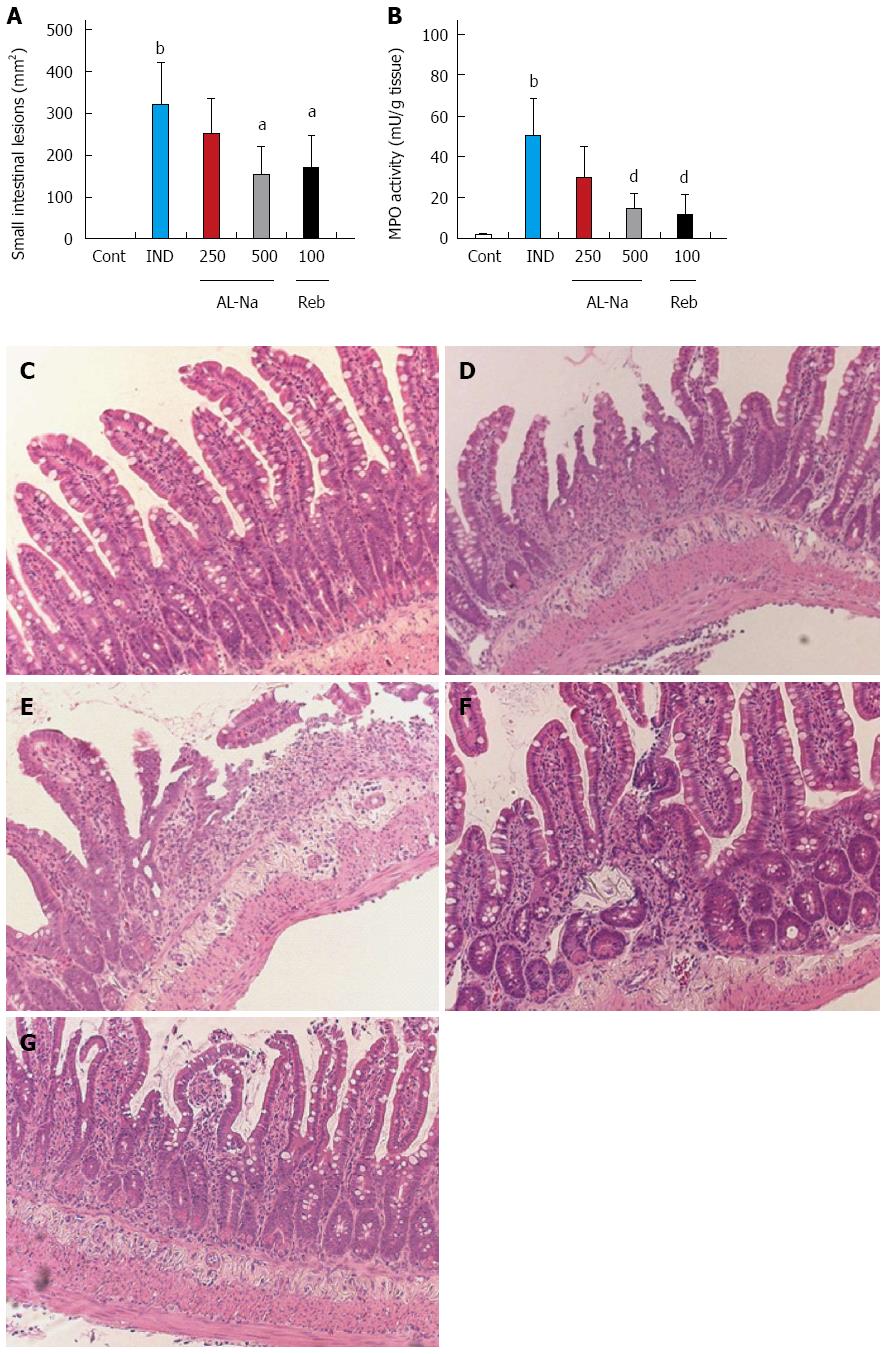
Figure 2 Effects of drugs on indomethacin-induced small intestinal lesions.
Animals were given indomethacin (10 mg/kg, po) and killed 24 h later. AL-Na (250 and 500 mg/kg) or Reb (100 mg/kg) was given orally twice at 30 min before and 6 h after administration of indomethacin. (A) The lesion areas and (B) myeloperoxidase (MPO) activity were measured. C: Hematoxylin and eosin-stained microscopic observations of the rat small intestinal mucosa of the control (Cont) group; D: Indomethacin (IND); E: AL-Na (250 mg/kg); F: AL-Na (500 mg/kg); G: Reb (100 mg/kg). Each column and vertical bar represents the mean ± SE (n = 8). Significantly different from the cont group at bP < 0.01 (Student’s t-test). Significantly different from the IND group at aP < 0.05 and dP < 0.01, respectively (Dunnett’s test). AL-Na: Sodium alginate; Reb: Rebamipide.
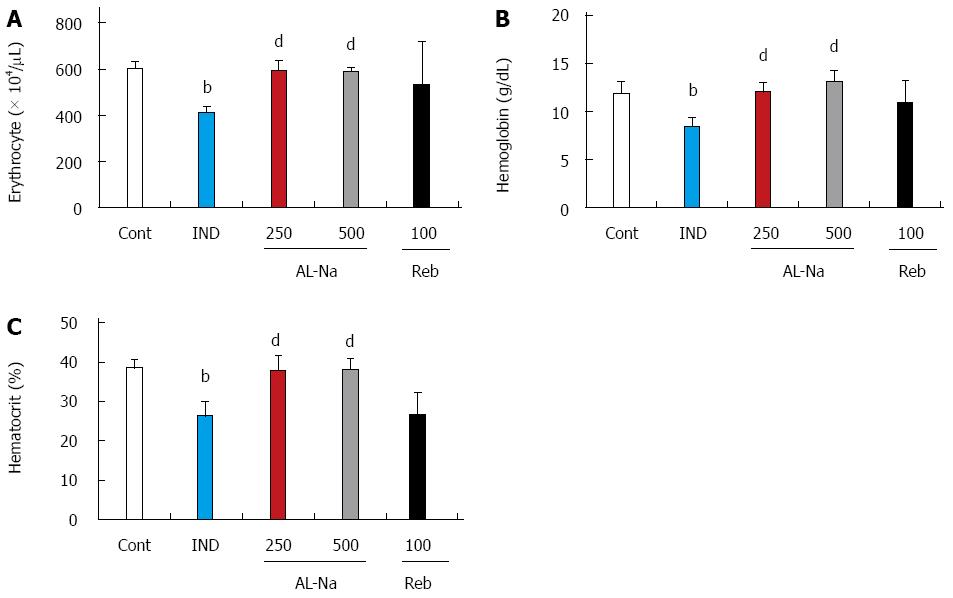
Figure 3 Effects of drugs on indomethacin-induced anemia.
Animals were given indomethacin (10 mg/kg, po), and blood samples were obtained 24 h later. AL-Na (250 and 500 mg/kg) or Reb (100 mg/kg) was given orally twice at 30 min before and 6 h after administration of indomethacin. A: Erythrocyte; B: Haemoglobin; C: Haematocrit. Each column and vertical bar represents the mean ± SE (n = 8). Significantly different from the control group at bP < 0.01 (Student’s t-test). Significantly different from the indomethacin group at dP < 0.01 (Dunnett’s test). Cont: Control; IND: Indomethacin; AL-Na: Sodium alginate; Reb: Rebamipide.
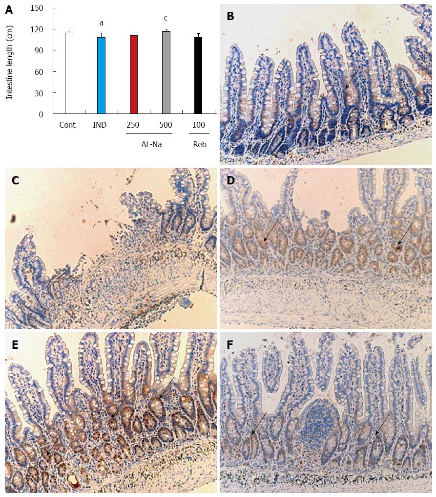
Figure 4 Effects of drugs on indomethacin-induced atrophy.
Animals were given indomethacin (10 mg/kg, po) and killed 24 h later. AL-Na (250 and 500 mg/kg) or Reb (100 mg/kg) was given orally twice at 30 min before and 6 h after administration of indomethacin. A: The small intestinal length was measured; B: Microscopic observations with proliferating cell nuclear antigen (PCNA) positive cells of the rat ileal mucosa of the control (Cont) group; C: Indomethacin (IND); D: AL-Na (250 mg/kg); E: AL-Na (500 mg/kg); F: Reb (100 mg/kg). Each column and vertical bar represents the mean ± SE (n = 8). Significantly different from the Cont group at aP < 0.05 (Student’s t-test); Significantly different from the IND group at cP < 0.05 (Dunnett’s test). AL-Na: Sodium alginate; Reb: Rebamipide. The allows show PCNA positive cells.
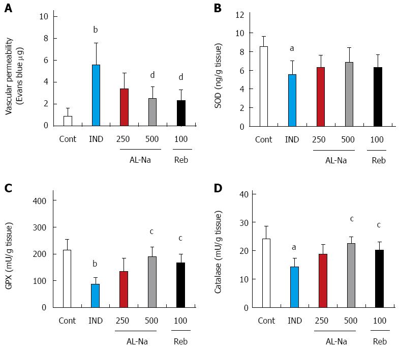
Figure 5 Effects of drugs on indomethacin-induced vascular permeability and oxidative stress.
Animals were given indomethacin (10 mg/kg, po) and killed 24 h later. AL-Na (250 and 500 mg/kg) or Reb (100 mg/kg) was given orally twice at 30 min before and 6 h after administration of indomethacin. A Vascular permeability; B: Superdismdeoxidase content; C: Glutathione peroxidase activity; D: Catalase activity were measured. Each column and vertical bar represents the mean ± SE (n = 8). Significantly different from the control group at aP < 0.05 and bP < 0.01 (Student’s t-test); Significantly different from the indomethacin group at cP < 0.05 and dP < 0.01, respectively (Dunnett’s test). Cont: Control; IND: Indomethacin; AL-Na: Sodium alginate; Reb: Rebamipide.
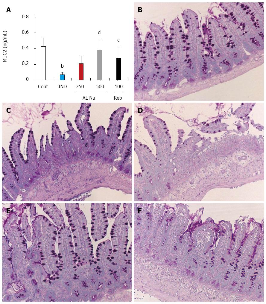
Figure 6 Effects of drugs on indomethacin-induced mucin depletion in small intestine.
Animals were given indomethacin (10 mg/kg, po) and killed 24 h later. AL-Na (250 and 500 mg/kg) or Reb (100 mg/kg) was given orally twice at 30 min before and 6 h after administration of indomethacin. A: The mucin of the small intestine was measured; B: PAS-stained microscopic observations of the rat small intestinal mucosa of the control (Cont) group; C: Indomethacin (IND); D: AL-Na (250 mg/kg); E: AL-Na (500 mg/kg); F: Reb (100 mg/kg). Each column and vertical bar represents the mean ± SE (n = 8). Significantly different from the Cont group at bP < 0.01 (Student’s t-test). Significantly different from the IND group at dP < 0.01 and cP < 0.05 (Dunnett’s test). AL-Na: Sodium alginate; Reb: Rebamipide.
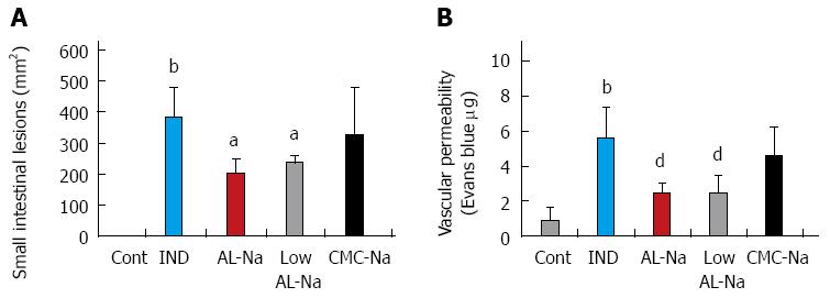
Figure 7 Effects of low molecular sodium alginate on indomethacin-induced small intestinal lesions.
Animals were given indomethacin (10 mg/kg, po) and killed 24 h later. Original AL-Na (500 mg/kg), low AL-Na (500 mg/kg) or CMC-Na (250 mg/kg) was given orally twice at 30 min before and 6 h after administration of indomethacin. (A) The lesion areas were measured. (B) The vascular permeability was measured. Each column and vertical bar represents the mean ± SE (n = 8). Significantly different from the control group at bP < 0.01 (Student’s t-test). Significantly different from the indomethacin group at aP < 0.05 and dP < 0.01 (Dunnett’s test). Cont: Control; IND: Indomethacin; AL-Na: Sodium alginate; Low AL-Na: Low molecular sodium alginate; CMC-Na: Sodium carboxymethylcellulose.
-
Citation: Yamamoto A, Itoh T, Nasu R, Nishida R. Sodium alginate ameliorates indomethacin-induced gastrointestinal mucosal injury
via inhibiting translocation in rats. World J Gastroenterol 2014; 20(10): 2641-2652 - URL: https://www.wjgnet.com/1007-9327/full/v20/i10/2641.htm
- DOI: https://dx.doi.org/10.3748/wjg.v20.i10.2641


