©The Author(s) 1996.
World J Gastroenterol. Sep 15, 1996; 2(3): 155-157
Published online Sep 15, 1996. doi: 10.3748/wjg.v2.i3.155
Published online Sep 15, 1996. doi: 10.3748/wjg.v2.i3.155
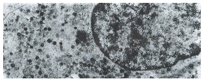
Figure 1 G cell of the antrum containing both vesicular and compact granules with a floccular content.
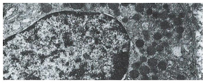
Figure 2 D cell granules are usually rounded, homogeneous, of low electron density and with closely applied membranes.
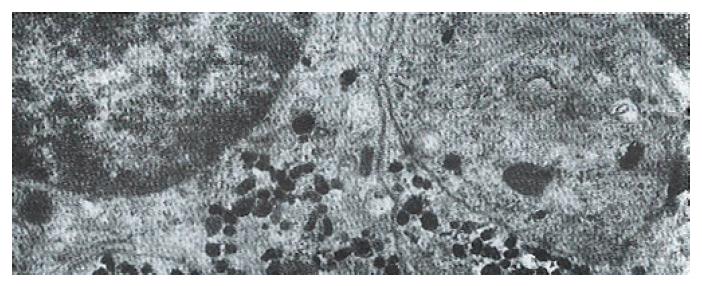
Figure 3 EC cell granules appear to be pleomorphic, such as being oblong, ovoid, round, etc.
, with high density.
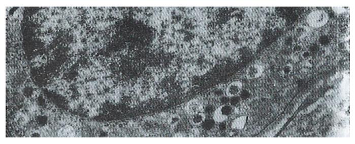
Figure 4 ECL cells show the characteristic of vesicular-type granules and thin haloed granules with an irregular argyrophilic core eccentrically located in wide space.
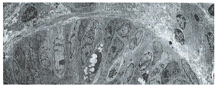
Figure 5 Electron micrograph of contiguous gastric glands shows cross sections belonging to different endocrine cells.
- Citation: Yu JY, Adda T. Quantitative ultrastructure analysis of neuroendocrine cells of gastric mucosa in normal and pathological conditions. World J Gastroenterol 1996; 2(3): 155-157
- URL: https://www.wjgnet.com/1007-9327/full/v2/i3/155.htm
- DOI: https://dx.doi.org/10.3748/wjg.v2.i3.155


