Copyright
©2013 Baishideng Publishing Group Co.
World J Gastroenterol. Mar 7, 2013; 19(9): 1489-1493
Published online Mar 7, 2013. doi: 10.3748/wjg.v19.i9.1489
Published online Mar 7, 2013. doi: 10.3748/wjg.v19.i9.1489
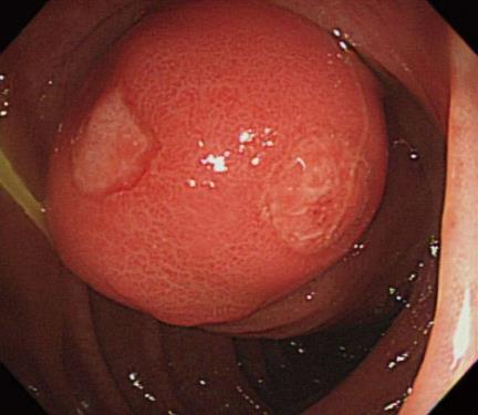
Figure 1 Colonoscopy disclosed a submucosal tumor over the cecum.
The mass was a ball-like form with eroded surface.
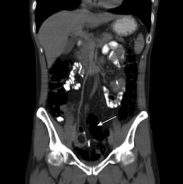
Figure 2 Computed tomography scan showing a round and low density mass about 2.
5 cm in the right pelvic region.
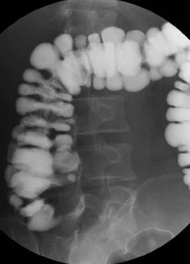
Figure 3 Barium enema study showed a bulging lesion over the cecum.
Figure 4 The terminal ileum had invaginated through the ileocecal valve and ileocolic intussusception was observed.
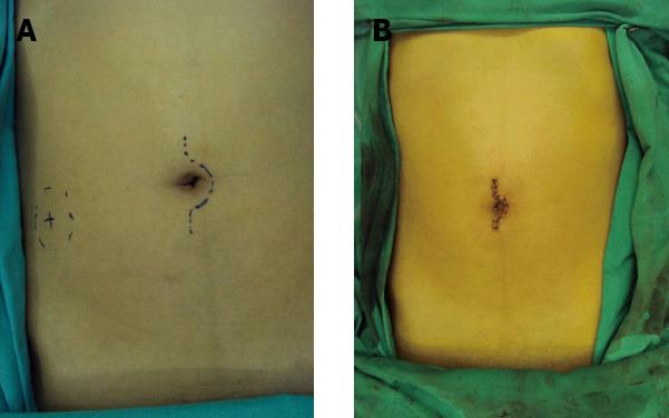
Figure 5 Single-port laparoscopic approach.
A: A vertical incision was created through the umbilicus approximately 3 cm in length which accommodated the single-port access device; B: Postoperative suture wound.
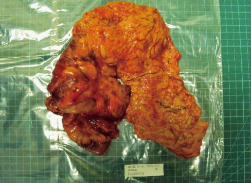
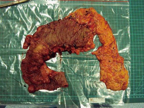
Figure 6 The round tumor measured 2.
5 cm × 2.5 cm × 2.0 cm in size and was 15 cm from the ileocecal valve.
- Citation: Chen JH, Wu JS. Single port laparoscopic right hemicolectomy for ileocolic intussusception. World J Gastroenterol 2013; 19(9): 1489-1493
- URL: https://www.wjgnet.com/1007-9327/full/v19/i9/1489.htm
- DOI: https://dx.doi.org/10.3748/wjg.v19.i9.1489


