Copyright
©2013 Baishideng Publishing Group Co.
World J Gastroenterol. Feb 21, 2013; 19(7): 1143-1146
Published online Feb 21, 2013. doi: 10.3748/wjg.v19.i7.1143
Published online Feb 21, 2013. doi: 10.3748/wjg.v19.i7.1143
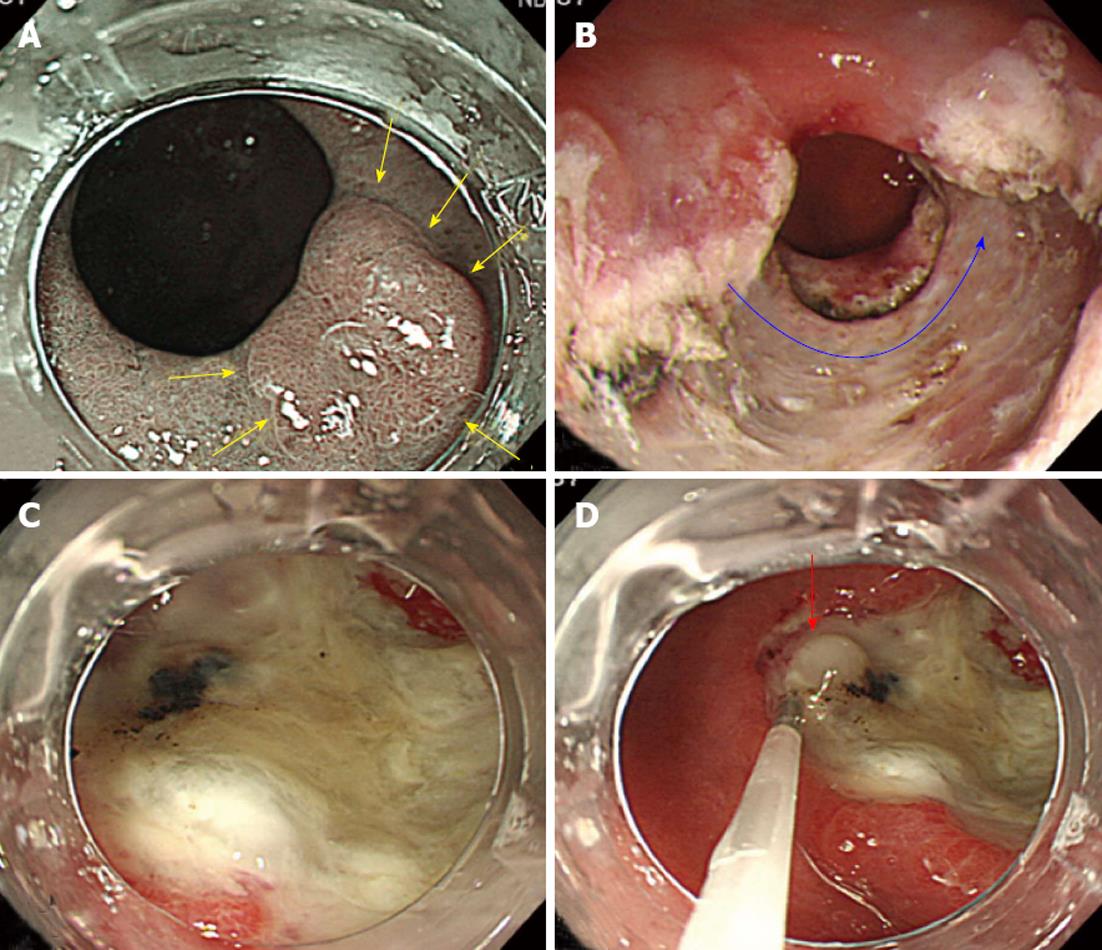
Figure 1 Endoscopic findings of tumor, post-endoscopic submucosal dissection ulcer and triamcinolone acetonide injection.
A: An narrow band imaging endoscopic image reveals a flat, early gastric cancer lesion extending over the gastric outlet to the pylorus (yellow arrows); B: A post-endoscopic submucosal dissection artificial ulcer covering two-thirds of the circumference of the pylorus (blue curved arrow); C: The ulcer floor covered by a thick layer of white moss; D: Triamcinolone acetonide (2 mL) was injected locally at each site (red arrow).
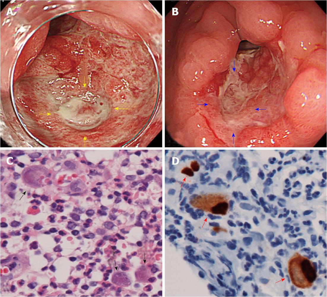
Figure 2 Endoscopic findings of cytomegalovirus associated ulcer and microscopic examination.
A: Artificial ulcer on postoperative day 12, showing the formation of abundant granulation tissue and a 10-mm-deep ulcer at the center of the granulation tissue (yellow arrows); B: The healing process of the deep ulcer (blue arrows) on postoperative day 15; C: A biopsy from the deeper ulcer margin revealed large cells with intranuclear inclusion bodies (black arrow, HE staining, × 600); D: Large cells with intranuclear inclusion bodies stained positive for anti-cytomegalovirus (CMV) antibodies (red arrows, anti-CMV antibody immunohistochemical staining, × 600).
- Citation: Mori H, Fujihara S, Nishiyama N, Kobara H, Oryu M, Kato K, Rafiq K, Masaki T. Cytomegalovirus-associated gastric ulcer: A side effect of steroid injections for pyloric stenosis. World J Gastroenterol 2013; 19(7): 1143-1146
- URL: https://www.wjgnet.com/1007-9327/full/v19/i7/1143.htm
- DOI: https://dx.doi.org/10.3748/wjg.v19.i7.1143


