Copyright
©2013 Baishideng Publishing Group Co.
World J Gastroenterol. Feb 21, 2013; 19(7): 1049-1055
Published online Feb 21, 2013. doi: 10.3748/wjg.v19.i7.1049
Published online Feb 21, 2013. doi: 10.3748/wjg.v19.i7.1049
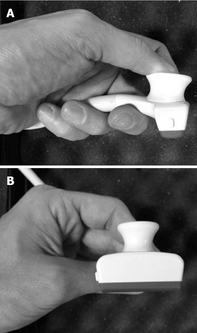
Figure 1 New probe for intraoperative ultrasound.
This probe has a trapezoid scanning area, and an ergonomic shape, which help during intraoperative ultrasound-guided maneuvers. A: Lateral view; B: Front view.
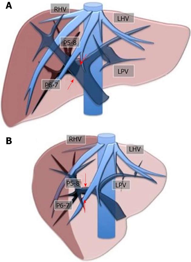
Figure 2 Layout of the liver for the inflow modulation.
Ischemic demarcation of the right posterior sector by intraoperative ultrasound-guided finger compression at its origin of the right portal bifurcation. A: Front view; B: Lateral view. RHV: Right hepatic vein; LHV: Left hepatic vein; LPV: Left portal vein; P5-8: Right anterior portal braches; P6-7: Right posterior portal branches. The arrows indicate the point for the compression.
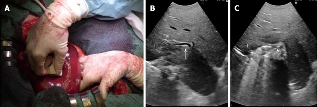
Figure 3 A case of intraoperative ultrasound-guided finger compression of segment 6.
A: The portal pedicle for segment 6 is compressed by the probe in the right hand and by the finger in the left hand; B: Intraoperative ultrasound (IOUS) focused on the portal pedicle (arrow) for segment 6 before the compression; C: IOUS focused on the portal pedicle (arrow) for segment 6 during the compression. T: Tumor.
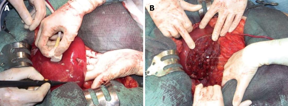
Figure 4 Demarcation of the compressed area by electrocautery.
A: The operative field before the resection; B: The operative field at the end of the resection. The arrow indicates the stump of the portal pedicle for segment 6.
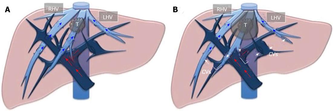
Figure 5 Layout of the liver for outflow modulation.
A: A tumor in contact with the middle hepatic vein at the caval confluence; B: Once that vein is infiltrated and/or compressed, some collateral veins (CVs) shunting the flow from the middle hepatic vein territory to right hepatic vein (RHV) and/or left hepatic vein (LHV) territories can be detected. T: Tumor.
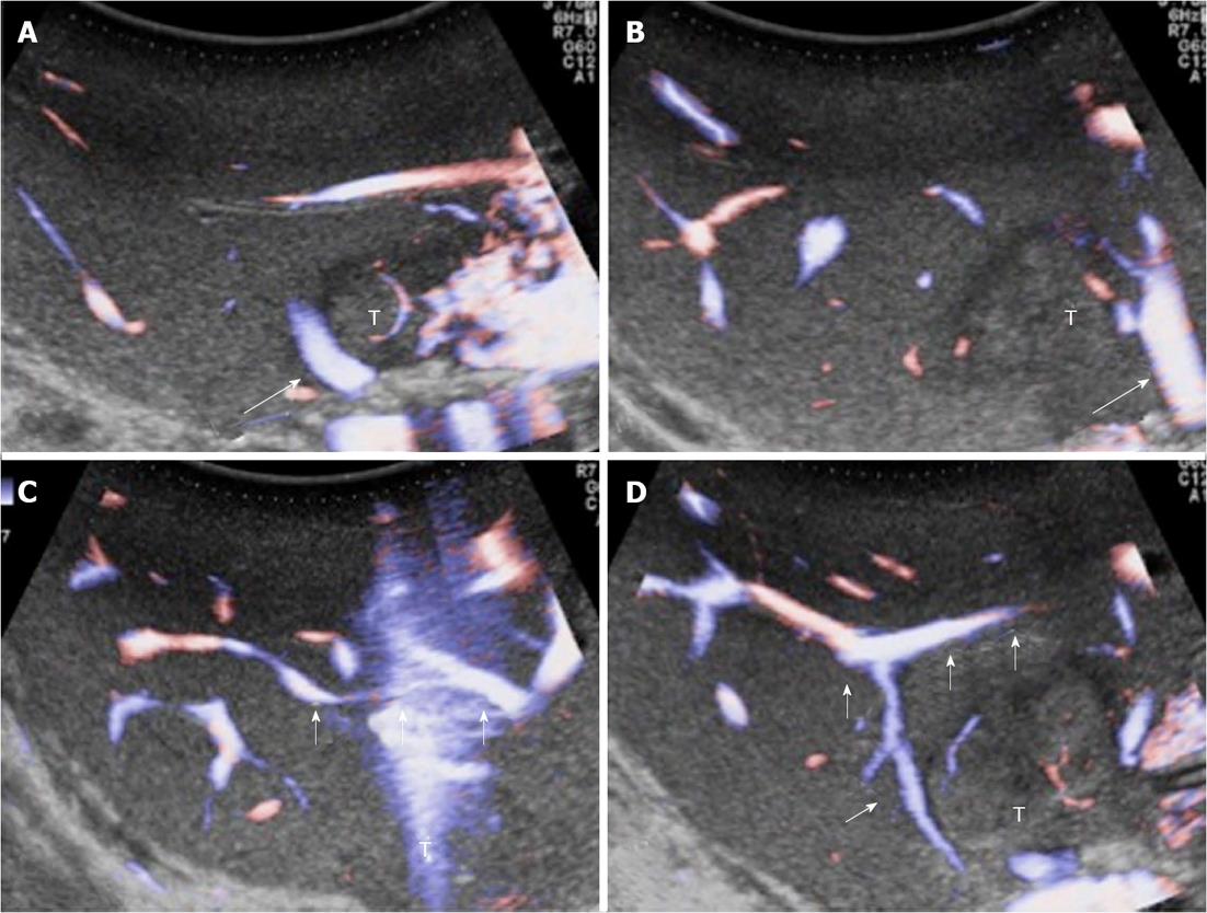
Figure 6 Intraoperative ultrasound study of communicating veins.
A: A tumour located between the middle hepatic vein (MHV) (arrow) and the left hepatic vein (LHV) at their confluence into the inferior vena cava; B: The arrow indicates the LHV; C, D: Evidence of communicating veins (arrows) between the LHV and the MHV. T: Tumor.
- Citation: Donadon M, Procopio F, Torzilli G. Tailoring the area of hepatic resection using inflow and outflow modulation. World J Gastroenterol 2013; 19(7): 1049-1055
- URL: https://www.wjgnet.com/1007-9327/full/v19/i7/1049.htm
- DOI: https://dx.doi.org/10.3748/wjg.v19.i7.1049


