©2013 Baishideng Publishing Group Co.
World J Gastroenterol. Feb 14, 2013; 19(6): 960-963
Published online Feb 14, 2013. doi: 10.3748/wjg.v19.i6.960
Published online Feb 14, 2013. doi: 10.3748/wjg.v19.i6.960
Figure 1 Abdominal computed tomographic images before surgery.
A: Non-contrast-enhanced computed tomography scan showing the lesion (arrow) located in the right lobe of the liver; B: The lesion (arrow) showed enhancement during the hepatic arterial phase; C: The lesion (arrow) showed no enhancement during the portal phase.
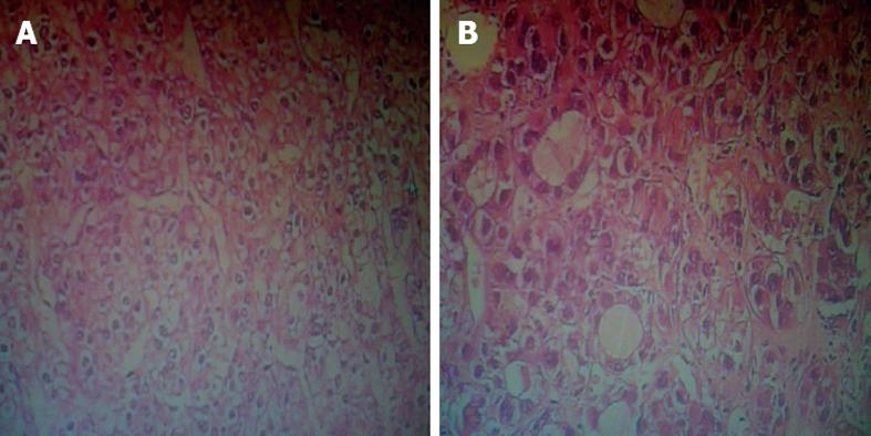
Figure 2 Histopathology of the primary tumor and enlarged left supraclavicular lymph node.
A: Cancer cells having oval and single nuclei, with less mitosis and less heteromorphosis in the primary lesion; hematoxylin and eosin (HE) stain, ×400; B: The same changes in cell morphology in left supraclavicular lymph node; HE stain, ×400.
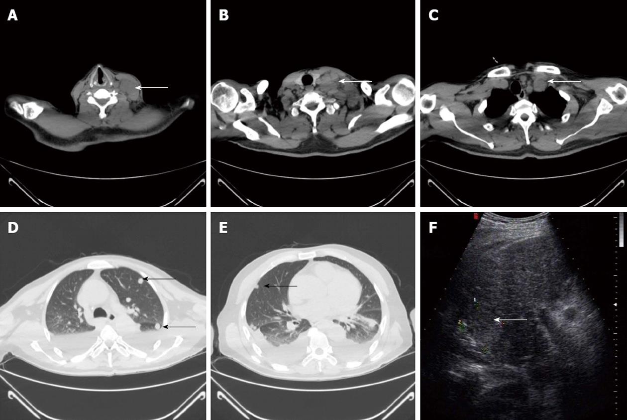
Figure 3 Thoracic computed tomographic and Doppler images after surgery.
A: Non-contrast-enhanced computed tomography scan showing enlarged left supraclavicular lymph nodes (arrow), which were easily observed; B: Part of enlarged left supraclavicular lymph nodes (arrow) fused into the mass; C: Enlarged lymph nodes in the mediastinum (arrow) was observed; D and E: Round nodules (arrows) distributed in both lungs; F: A new intrahepatic lesion (arrow) was detected in the non-operating region via Doppler ultrasound.
- Citation: Liu T, Gao JF, Yi YX, Ding H, Liu W. Misdiagnosis of left supraclavicular lymph node metastasis of hepatocellular carcinoma: A case report. World J Gastroenterol 2013; 19(6): 960-963
- URL: https://www.wjgnet.com/1007-9327/full/v19/i6/960.htm
- DOI: https://dx.doi.org/10.3748/wjg.v19.i6.960


