©2013 Baishideng Publishing Group Co.
World J Gastroenterol. Nov 28, 2013; 19(44): 8146-8150
Published online Nov 28, 2013. doi: 10.3748/wjg.v19.i44.8146
Published online Nov 28, 2013. doi: 10.3748/wjg.v19.i44.8146
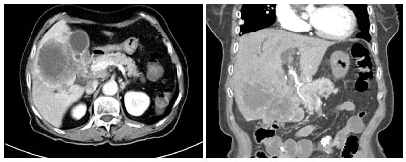
Figure 1 Findings of initial abdominal computed tomography.
Computed tomography showed a large mass located in the liver, common hepatic duct, and common bile duct.
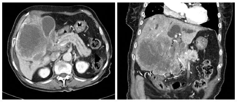
Figure 2 Computed tomography scan obtained before biopsy.
One month after the first visit, a computed tomography scan of the pancreas and biliary duct revealed that the large mass in the right lobe of the liver had grown and that there was an obstruction of the hilar duct.
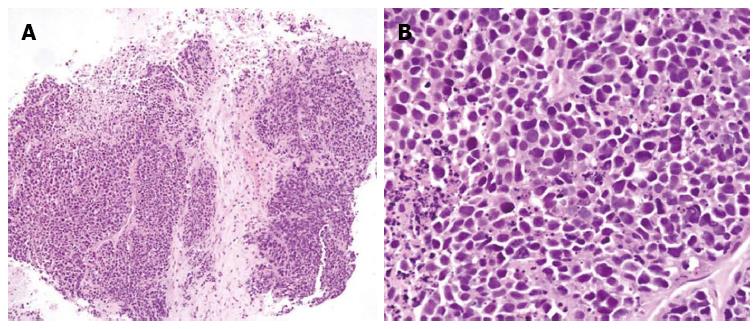
Figure 3 Tumor consisted of tightly packed nests and diffuse, irregularly shaped sheets of cells with necrotic areas.
A: The tumor cells were of small-to-intermediate size with hyperchromatic, round-to-oval nuclei and scanty, poorly defined cytoplasm (HE, × 100); B: The nuclear chromatin was finely granular, and nucleoli were absent or inconspicuous. Cell borders were rarely seen, and nuclear molding was common (HE, × 400).
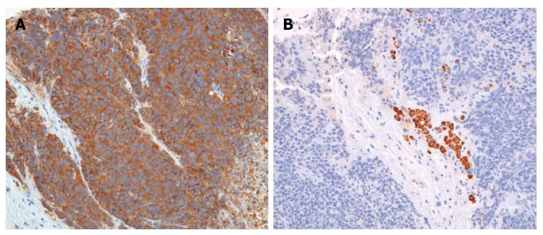
Figure 4 Immunohistochemical findings (× 200).
The tumor cells were immunoreactive for synaptophysin (A) and negative for hepatocyte-specific antigen (B).
- Citation: Jo JM, Cho YK, Hyun CL, Han KH, Rhee JY, Kwon JM, Kim WK, Han SH. Small cell carcinoma of the liver and biliary tract without jaundice. World J Gastroenterol 2013; 19(44): 8146-8150
- URL: https://www.wjgnet.com/1007-9327/full/v19/i44/8146.htm
- DOI: https://dx.doi.org/10.3748/wjg.v19.i44.8146


