Copyright
©2013 Baishideng Publishing Group Co.
World J Gastroenterol. Nov 21, 2013; 19(43): 7696-7700
Published online Nov 21, 2013. doi: 10.3748/wjg.v19.i43.7696
Published online Nov 21, 2013. doi: 10.3748/wjg.v19.i43.7696
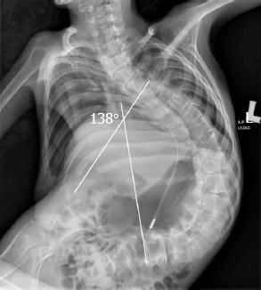
Figure 1 A 17-year-old patient with severe kyphoscoliosis (138 degrees on Cobb’s scale) with interposed organs.
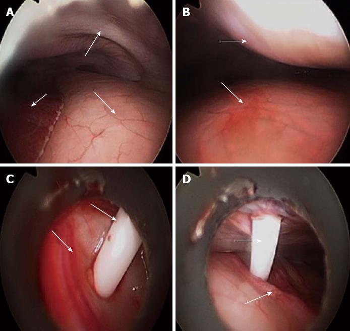
Figure 2 Laparoscopy-assisted percutaneous endoscopic gastrostomy placement: An overview of the procedure.
A: General view. The arrows indicate the following (respectively): liver, stomach and abdominal wall; B: Marking the puncture site. The arrows indicate the stomach and abdominal walls, and the pressed wall shows the puncture site; C: Introduction of the needle into the stomach under visual control. The arrows show the gastric wall and the trocar for percutaneous endoscopic gastrostomy (PEG) insertion; D: PEG was performed. The arrows indicate the gastric vessels and the trocar.
- Citation: Hermanowicz A, Matuszczak E, Komarowska M, Jarocka-Cyrta E, Wojnar J, Debek W, Matysiak K, Klek S. Laparoscopy-assisted percutaneous endoscopic gastrostomy enables enteral nutrition even in patients with distorted anatomy. World J Gastroenterol 2013; 19(43): 7696-7700
- URL: https://www.wjgnet.com/1007-9327/full/v19/i43/7696.htm
- DOI: https://dx.doi.org/10.3748/wjg.v19.i43.7696


