Copyright
©2013 Baishideng Publishing Group Co.
World J Gastroenterol. Nov 21, 2013; 19(43): 7544-7551
Published online Nov 21, 2013. doi: 10.3748/wjg.v19.i43.7544
Published online Nov 21, 2013. doi: 10.3748/wjg.v19.i43.7544
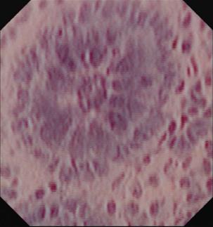
Figure 1 Endocytoscopic image at magnification × 1390 showing one single colonic crypt.
Goblet and epithelial cells are clearly evident.
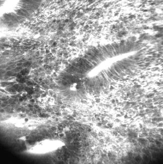
Figure 2 Endoscope-based confocal laser endomicroscopy of Crohn’s disease shows remarkable cell infiltration, disturbed colonic architecture and leakage indicating severe inflammation.
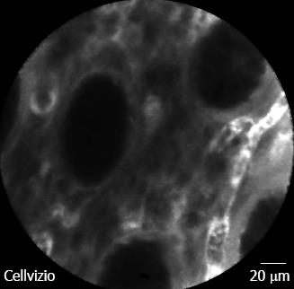
Figure 3 Probe-based confocal laser endomicroscopy in patient with Crohn’s disease without histologic activity.
Colonic crypts are regularly arranged with normal shape and size. Micro-vessels within the lamina propria are easily visible. Bar = 20 μm.
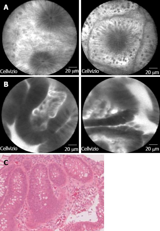
Figure 4 Probe-based confocal laser endomicroscopy images of (A) normal colonic mucosa; B: Adenomatous polyp with low grade dysplasia.
Bar = 20 mm; and C: The corresponding histologic image for the adenomatous polyp with dysplasia (magnification × 40).
- Citation: Liu J, Dlugosz A, Neumann H. Beyond white light endoscopy: The role of optical biopsy in inflammatory bowel disease. World J Gastroenterol 2013; 19(43): 7544-7551
- URL: https://www.wjgnet.com/1007-9327/full/v19/i43/7544.htm
- DOI: https://dx.doi.org/10.3748/wjg.v19.i43.7544


