©2013 Baishideng Publishing Group Co.
World J Gastroenterol. Nov 7, 2013; 19(41): 7032-7041
Published online Nov 7, 2013. doi: 10.3748/wjg.v19.i41.7032
Published online Nov 7, 2013. doi: 10.3748/wjg.v19.i41.7032
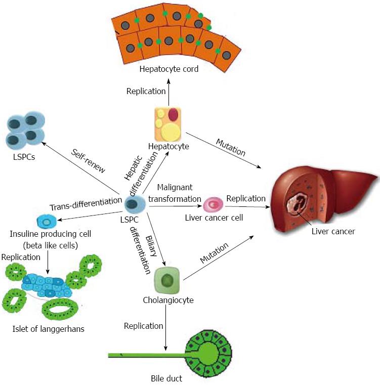
Figure 1 The different cell fates of liver stem/progenitor cells under distinct situations.
Under special stem microenviroment, LSPCs would probably self renew to keep stem properties. In the contrary, under differentiating stimuli, LSPCs could give birth to two kinds of fundermental mature cells in the liver, hepatocytes and cholangiocytes. This is very important for liver development and liver regeneration. Except for the traditional differentiation directions, LSPCs have the capacity to trans-differentiate into insulin producing cells, which is promising for treating diabetes. While in some bad situations, LSPCs may carry out malignant transformation to become liver cancer cells, even liver cancer stem cells, as a result, these cancer cells replicate themselves to cause liver cancer. This is a different way from mutation of hepatocytes for liver carcinogenesis. LSPC: Liver stem/progenitor cell.
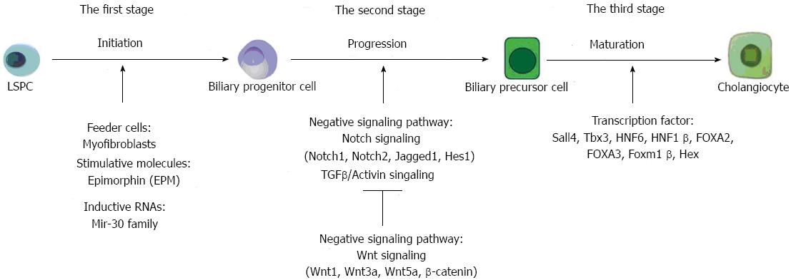
Figure 2 The molecular mechanisms in each step of biliary differentiation of liver stem/progenitor cells.
The biliary differentiation of LSPCs can be divided into three main stages comprising of initiation, progression and maturation. Some important jacent or feeder cells and specific molecules are proposed to be responsible for initiating the first stage of biliary differentiation. When the biliary differentiation goes on, several key signaling pathways including Notch and TGFβ have essential impacts on guarantee of the second stage. After some transcription factors activated, the third stage of biliary differentiation could be accomplished and LSPCs could be matured into cholangiocytes. LSPCs: Liver stem/progenitor cells; TGFβ: Transforming growth factor β.
- Citation: Liu WH, Ren LN, Chen T, Liu LY, Tang LJ. Stages based molecular mechanisms for generating cholangiocytes from liver stem/progenitor cells. World J Gastroenterol 2013; 19(41): 7032-7041
- URL: https://www.wjgnet.com/1007-9327/full/v19/i41/7032.htm
- DOI: https://dx.doi.org/10.3748/wjg.v19.i41.7032


