Copyright
©2013 Baishideng Publishing Group Co.
World J Gastroenterol. Oct 14, 2013; 19(38): 6494-6499
Published online Oct 14, 2013. doi: 10.3748/wjg.v19.i38.6494
Published online Oct 14, 2013. doi: 10.3748/wjg.v19.i38.6494
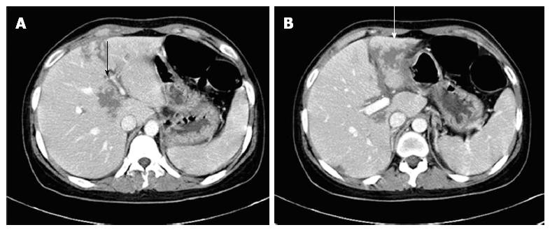
Figure 1 Intraparenchymal hemorrhage in the liver (arrows).
An abdominal computed tomography showed multiple low attenuated lesions without peripheral enhancement (A) and perilesional edema of the liver on the contrast-enhanced image (B).
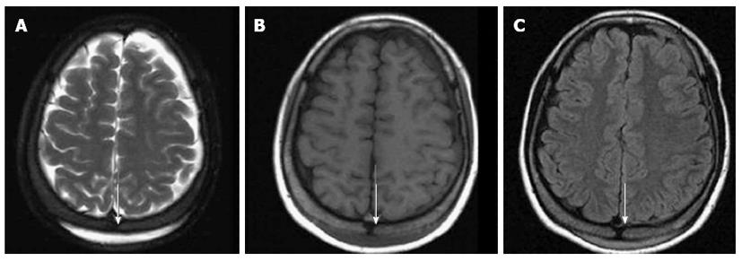
Figure 2 Subgaleal hematoma at the posterior parietal scalp (arrows).
A brain magnetic resonance imaging showed a crescentic high signal in the posterior parietal scalp on the T2 weighted image (A), a low signal on the T1 weighted image (B) and mild enhancement on the contrast-enhanced image (C).
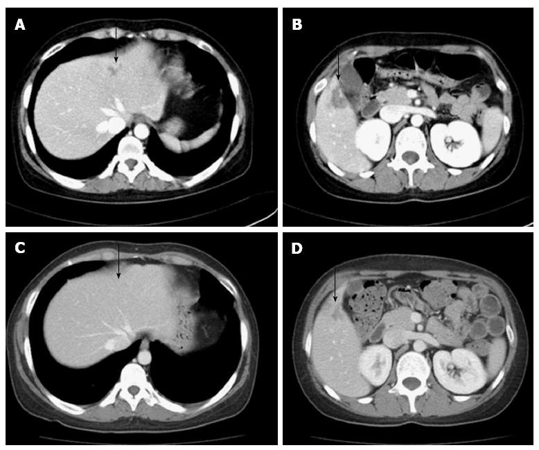
Figure 3 Recurrent and nearly complete resolution intraparenchymal hemorrhage in the liver (arrows).
A, B: An abdominal computed tomography showed a low attenuated lesion (arrow) in the S2/4 (A) and S5 segments (B) of the liver on the contrast-enhanced image; C, D: An abdominal computed tomography showed resolution of the parenchymal hemorrhage in S2/4 (C) and the healing process in S5 (D) of the liver.
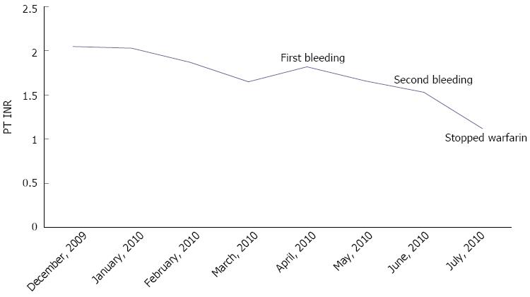
Figure 4 Extended follow-up data for the prothrombin time international normalized ratio.
PT INR: Prothrombin time international normalized ratio.
- Citation: Park IC, Baek YH, Han SY, Lee SW, Chung WT, Lee SW, Kang SH, Cho DS. Simultaneous intrahepatic and subgaleal hemorrhage in antiphospholipid syndrome following anticoagulation therapy. World J Gastroenterol 2013; 19(38): 6494-6499
- URL: https://www.wjgnet.com/1007-9327/full/v19/i38/6494.htm
- DOI: https://dx.doi.org/10.3748/wjg.v19.i38.6494


