©2013 Baishideng Publishing Group Co.
World J Gastroenterol. Jun 21, 2013; 19(23): 3693-3698
Published online Jun 21, 2013. doi: 10.3748/wjg.v19.i23.3693
Published online Jun 21, 2013. doi: 10.3748/wjg.v19.i23.3693
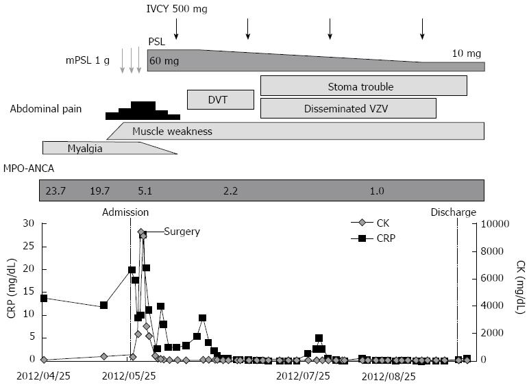
Figure 1 Clinical course.
CK: Creatinine kinase; CRP: C-reactive protein; DVT: Deep venous thrombosis; IVCY:Intravenous cyclophosphamide; mPSL:Methylprednisolone; MPO-ANCA:Myeloperoxidase-anti-neutrophil cytoplasmic antibody; PSL: Prednisolone; VZV: Varicella zoster virus.
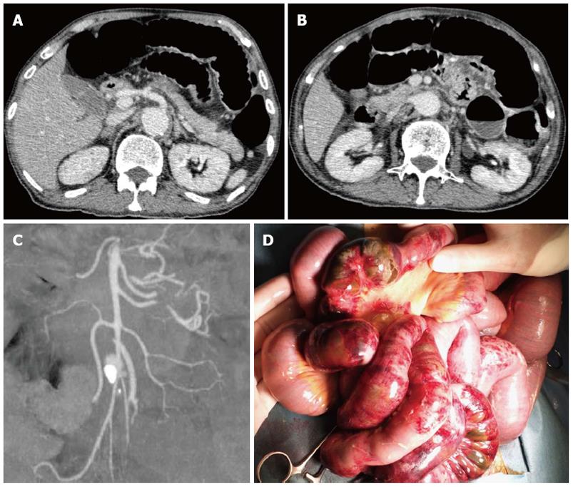
Figure 2 Clinical imaging.
A: Computed tomography (CT) image of the abdomen showing distended small bowel loops, gas in the small bowel, blurred enhancement of the intestinal wall, and absence of any significant obstruction in the celiac trunk; B: CT image of the abdomen showing superior mesenteric artery; C: CT angiographic reconstruction of the superior mesenteric artery; D: Surgical findings showing distended small intestine, segmental intestinal ischemia, and necrosis.
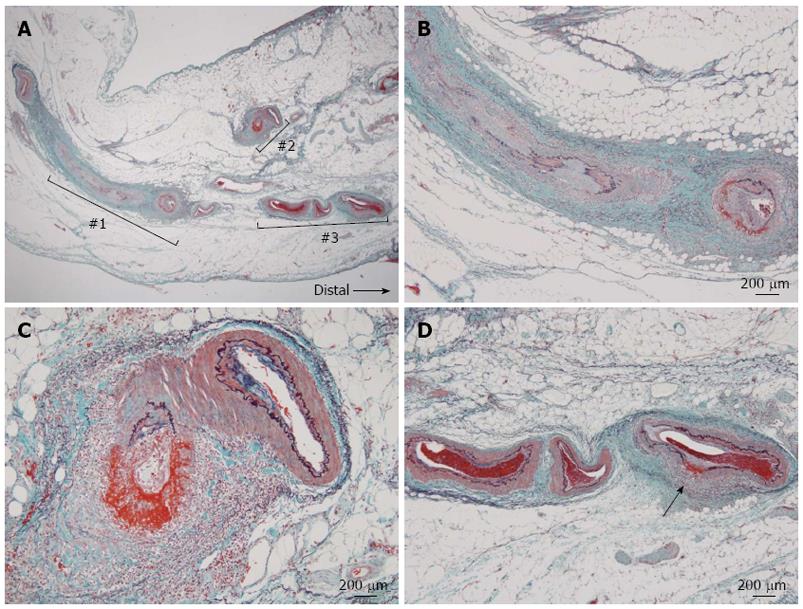
Figure 3 Histological findings of the jejunum.
A: Necrotizing vasculitis of the mesenteric artery showing nodular lesions in various stages of the Arkin classification, Elastica-Masson staining; B: Higher magnification of No. 1 in A showing necrotizing vasculitis of mesenteric artery in stage II-III ; C: Higher magnification of No. 2 in A showing necrotizing vasculitis of the mesenteric artery in stage II and pan-arterial necrosis with fibrinoid degeneration; D: Higher magnification of No. 3 in A showing necrotizing vasculitis of mesenteric artery in stage II, characterized by destruction of internal elastic lamina associated with fibrinoid necrosis (arrow).
- Citation: Shirai T, Fujii H, Saito S, Ishii T, Yamaya H, Miyagi S, Sekiguchi S, Kawagishi N, Nose M, Harigae H. Polyarteritis nodosa clinically mimicking nonocclusive mesenteric ischemia. World J Gastroenterol 2013; 19(23): 3693-3698
- URL: https://www.wjgnet.com/1007-9327/full/v19/i23/3693.htm
- DOI: https://dx.doi.org/10.3748/wjg.v19.i23.3693


