©2013 Baishideng Publishing Group Co.
World J Gastroenterol. Jun 21, 2013; 19(23): 3608-3614
Published online Jun 21, 2013. doi: 10.3748/wjg.v19.i23.3608
Published online Jun 21, 2013. doi: 10.3748/wjg.v19.i23.3608
Figure 1 Confirmation of clinical response on follow-up endoscopy before and after Kaposi’s sarcoma therapy.
A: Gastrointestinal Kaposi sarcoma (arrows) in the esophagogastric junction before Kaposi’s sarcoma (KS) therapy; B: After four months of KS therapy with liposomal anthracycline.
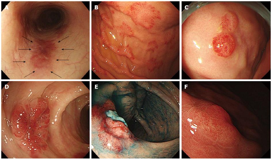
Figure 2 Gastrointestinal Kaposi sarcoma and mimicking lesions on endoscopy.
A: Kaposi sarcoma, dark reddish patch (arrows) in the esophagus; B: Kaposi sarcoma, multiple patch appearance in the stomach; C: Kaposi sarcoma, small (< 10 mm) and polypoid appearance in the stomach; D: Kaposi sarcoma, submucosal tumor (SMT) appearance with large size (≥ 10 mm) in the sigmoid colon; E: Kaposi sarcoma, submucosal tumor (SMT) appearance with ulceration in the ileo-cecal valve with indigo-carmine dye; F: Hyperplastic polyps mimicking Kaposi sarcoma with small size (< 10 mm) in the stomach.
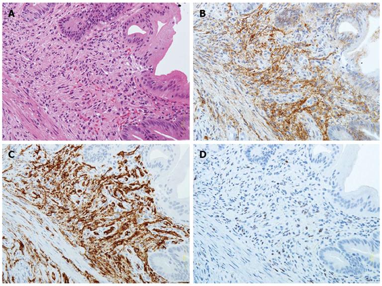
Figure 3 Pathological features of gastrointestinal Kaposi sarcoma.
A: Spindle cell proliferation in the submucosa on hematoxylin and eosin (HE) staining; B: Vascular gaps are lined with endothelial cells when staining with D2-40; C: Vascular gaps are lined with endothelial cells when staining with CD34; D: Some endothelial cells are positive for human herpesvirus 8.
- Citation: Nagata N, Igari T, Shimbo T, Sekine K, Akiyama J, Hamada Y, Yazaki H, Ohmagari N, Teruya K, Oka S, Uemura N. Diagnostic value of endothelial markers and HHV-8 staining in gastrointestinal Kaposi sarcoma and its difference in endoscopic tumor staging. World J Gastroenterol 2013; 19(23): 3608-3614
- URL: https://www.wjgnet.com/1007-9327/full/v19/i23/3608.htm
- DOI: https://dx.doi.org/10.3748/wjg.v19.i23.3608


