©2013 Baishideng Publishing Group Co.
World J Gastroenterol. May 28, 2013; 19(20): 3027-3042
Published online May 28, 2013. doi: 10.3748/wjg.v19.i20.3027
Published online May 28, 2013. doi: 10.3748/wjg.v19.i20.3027
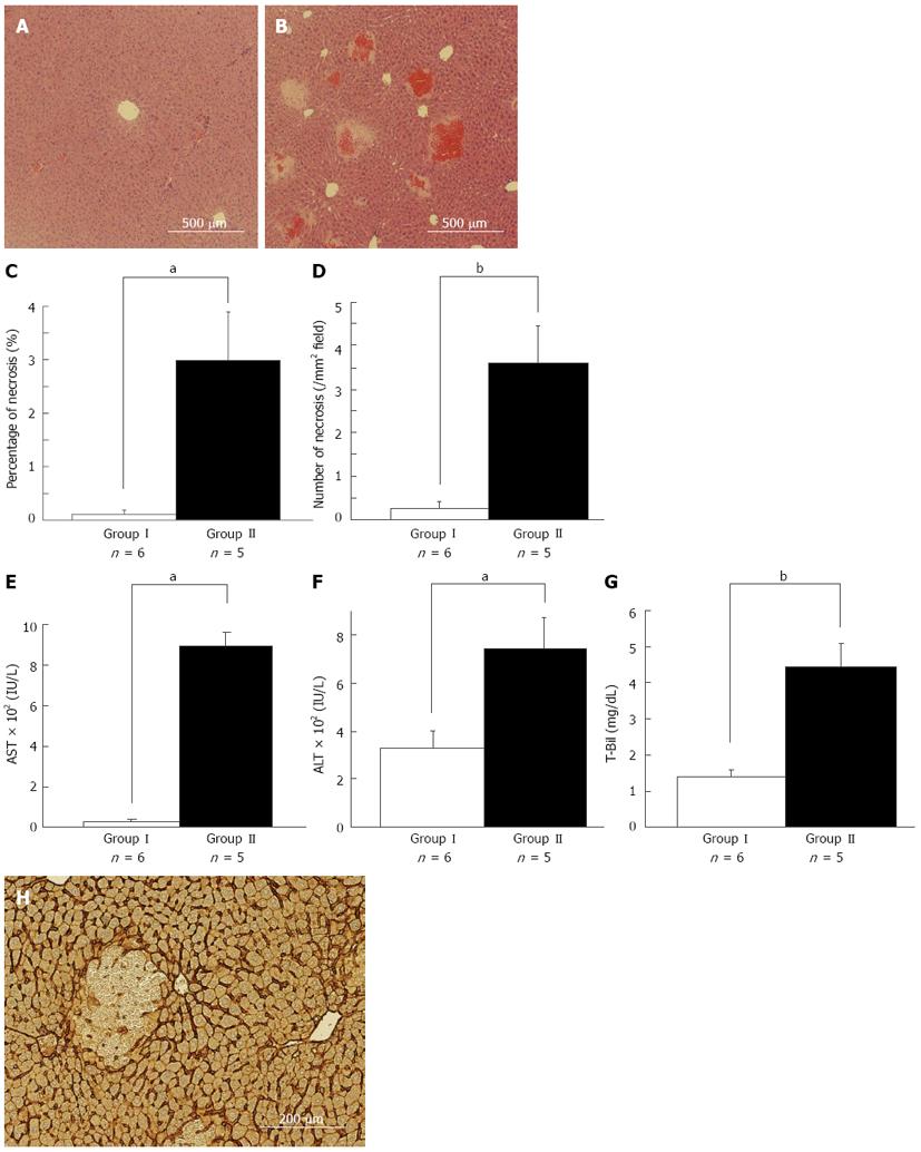
Figure 1 Necrosis is a pivotal event for postoperative liver failure in the remnant liver after 80%-partial hepatectomy.
There were histological and hematological differences between asymptomatic and symptomatic mice suspected as having postoperative liver failure 6 h after 80%-partial hepatectomy (PH) (A, B). Representative images of the remnant liver (RL) 6 h after 80%-PH in asymptomatic mice (A) and symptomatic mice (B) are shown. Significant multiple necroses with microhemorrhage were observed in the middle zone in symptomatic mice (B), while no obvious liver damage was observed in asymptomatic mice (A). The percentage area of necrosis (C) and the number of necrosis in each mm2 (D) in the RL were confirmed. Necrotic areas were significantly larger and more prevalent in symptomatic mice liver compared with asymptomatic mice. Serum levels of aspartate aminotransferase (AST) (E), alamine aminotransferase (ALT) (F) and total bilirubin (T-Bil) (G) 6 h after 80%-PH are shown. AST, ALT and T-Bil levels were significantly elevated in symptomatic mice compared with those in asymptomatic mice. Representative image of immunohistochemistry of intercellular adhesion molecule-1 (ICAM-1) in the RL 6 h after 80%-PH is shown (H). ICAM-1 expression was clearly observed, particularly in the sinusoid lining in areas without necrosis. We observed a breakdown in sinusoid structure with hemorrhage in necrotic areas after 80%-PH in mice, aP < 0.05, bP < 0.01 vs asymptomatic mice.
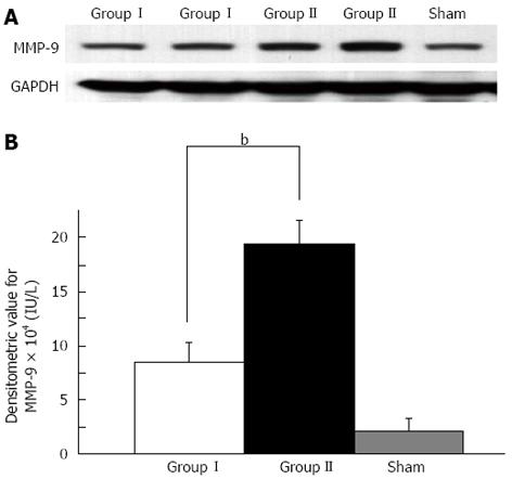
Figure 2 Western blotting analyses for matrix metalloproteinase-9 were performed in the remnant liver in asymptomatic mice and symptomatic mice after 80%-partial hepatectomy.
Representative image (A) and histograms (B) of Western blotting analyses for matrix metalloproteinase-9 (MMP-9) are shown. MMP-9 protein expression in the liver was enhanced in symptomatic mice in the process of postoperative liver failure compared with that in asymptomatic mice after extended hepatectomy and sham-treated mice (bP < 0.01 between Groups I and II).
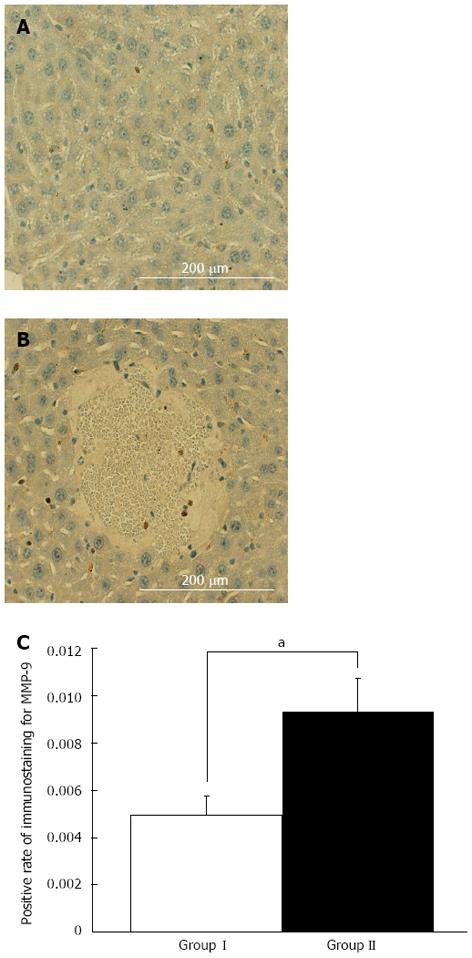
Figure 3 Immunohistochemical analysis for matrix metalloproteinase-9 in the remnant liver in symptomatic mice after 80%-partial hepatectomy was performed.
A: Representative images of matrix metalloproteinase-9 (MMP-9) staining in an area without necrosis; B: A necrotic area in the same sample are shown. Histograms of the results of distribution analysis show MMP-9 expression; C: Enhanced expression was observed in the areas close to necrosis compared with areas without necrosis in damaged RL (aP < 0.05 between Groups I and II).
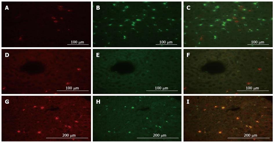
Figure 4 Localization analysis of matrix metalloproteinase-9 by dual immune-fluorescence in the remnant liver after 80%-partial hepatectomy is shown.
A: Fluorescent microscopy images of matrix metalloproteinase-9 (MMP-9) labeled in red (Alexa Fluor 568); B: CD68 labeled in green (Alexa Fluor 488); C: A merged image of MMP-9 and CD68 staining are shown; D: MMP-9 labeled in red (Alexa Fluor 568); E: Desmin labeled in green (Alexa Fluor 488); and F: A merged image of MMP-9 and desmin are shown; G: MMP-9 labeled in red (Alexa Fluor 568); H: CD11b labeled in green (Alexa Fluor 488); I: A merged image of MMP-9 and CD11b are shown. Protein expression of MMP-9 was mainly localized in CD11b-positive cells and desmin-positive cells to some degree.
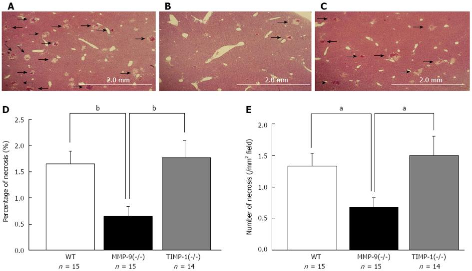
Figure 5 Histological analysis between wild-type, matrix metalloproteinase-9(-/-), and tissue inhibitor of metalloproteinase-1(-/-) mice 6 h after 80%-partial hepatectomy were performed.
Representative images of the remnant liver (RL) 6 h after 80%-PH in wild-type (WT) (A), matrix metalloproteinase-9 (MMP-9)(-/-) (B), and tissue inhibitor of metalloproteinase-1 (TIMP-1)(-/-) mice (C) are shown. Samples that included necrosis were selected for this figure. The percentage (D) and the number of necrotic foci per mm2 (E) of the necrotic area in the RL are shown. We observed significantly smaller and less necrotic foci in MMP-9(-/-) mice compared with WT and TIMP-1(-/-) mice (aP < 0.05, bP < 0.01 vs WT and TIMP-1).
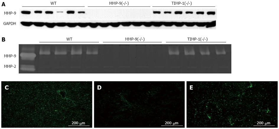
Figure 6 Matrix metalloproteinase-9 activity in the remnant liver with wild-type, matrix metalloproteinase-9(-/-), and tissue inhibitor of metalloprote-inase-1(-/-) mice 6 h after 80%-partial hepatectomy is shown.
A: Western blotting analysis for matrix metalloproteinase-9 (MMP-9) and glyceraldehyde 3-phosphate dehydrogenase (GAPDH) for a loading control was performed. The expression of MMP-9 in wild-type (WT) and tissue inhibitor of metalloproteinase-1 (TIMP-1)(-/-) mice and deletion in MMP-9(-/-) mice were observed at the protein level; B: Gelatin zymography analysis is shown; C-E: Pro-form MMP-9 activity and a slightly active form of MMP-9 were detected in WT and TIMP-1(-/-) mice. The activity of MMP-2 was the same depending on its genotype. In situ gelatin zymography analysis using DQ gelatin for gelatinolytic activity in the liver tissue is shown. A reduction in gelatinolytic activity in MMP-9(-/-) mice and enhancement in TIMP-1(-/-) mice are observed.
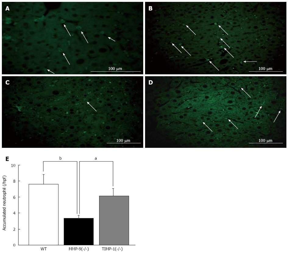
Figure 7 Immunohistochemistry analysis for accumulated neutrophils with myeloperoxidase staining between wild-type, matrix metalloproteinase-9(-/-) and tissue inhibitor of metalloprote-inase-1(-/-) mice 6 h after 80%-partial hepatectomy was performed.
Myeloperoxidase (MPO) staining labeled in green (Alexa Fluor 488) is shown (A). Cytoplasmic azurophilic granules were characteristically stained in neutrophils. Representative images of MPO staining on a remnant liver (RL) section in green in wild-type (WT) (B), matrix metalloproteinase-9 (MMP-9)(-/-) (C), tissue inhibitor of metalloproteinase-1 (TIMP-1)(-/-) mice (D) are shown. A histogram of the number of accumulated neutrophils in the liver is shown. We observed significantly fewer neutrophils in the RL in MMP-9(-/-) mice than in WT and TIMP-1(-/-) mice (aP < 0.05, bP < 0.01 vs WT and TIMP-1) (E).
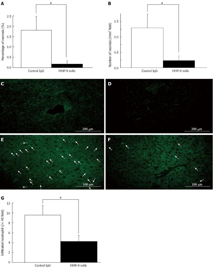
Figure 8 Inhibition of matrix metalloproteinase-9 by a monoclonal antibody of matrix metalloproteinase-9.
We investigated the outcome of inhibition of matrix metalloproteinase-9 (MMP-9) by monoclonal antibody (mAb) in 80%-partial hepatectomy (PH) in mice, and performed histological analysis of liver necrosis 6 h after 80%-PH (A, B). Liver damage was significantly reduced in the group with mAb compared with that in the control IgG group. Serum levels of aspartate aminotransferase, alamine aminotransferase and total bilirubin were lower in the mAb-treated mice, but this was not significant. Representative images of in situ gelatin zymography analysis with DQ gelatin revealed that gelatinolytic activity was suppressed in the mAb-treated mice (D) compared with that in control IgG-treated mice (C). Representative images of myeloperoxidase staining on remnant liver (RL) sections in control IgG-treated mice (E) and mAb-treated mice (F) are shown. A histogram of the number of accumulated neutrophils in the RL 6 h after 80%-PH shows that there are significantly fewer neutrophils in mAb-treated mice than in IgG-treated controls (G) (aP < 0.05 vs IgG group).
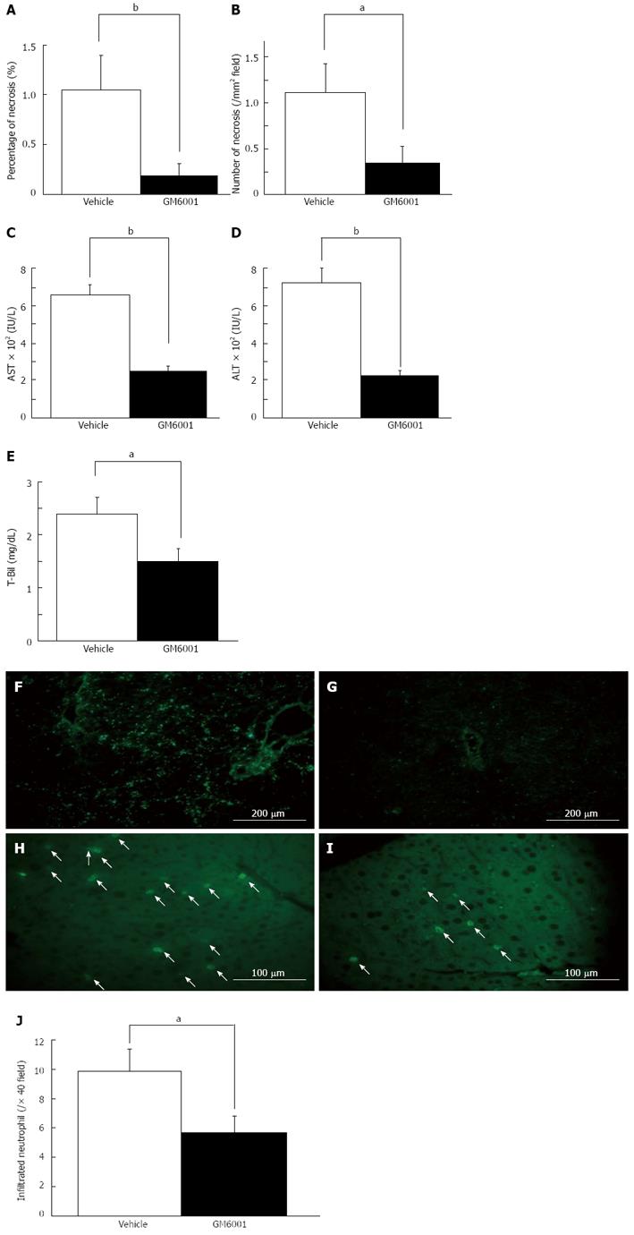
Figure 9 Broad-spectrum matrix metalloproteinase-9 inhibitor GM6001 ameliorate initial injury of the liver 6 h after 80%-partial hepatectomy.
We inhibited matrix metalloproteinase-9 by GM6001 in 80%-partial hepatectomy (PH) mice, and determined liver necrosis 6 h after 80%-PH. Liver damage was significantly reduced in the GM6001-treated mice compared with that in the vehicle-treated group (A, B). Serum levels of aspartate aminotransferase (AST) (C), alamine aminotransferase (ALT) (D) and total bilirubin (T-Bil) (E) were significantly lower in the GM6001-treated mice than those in the vehicle-treated mice. Representative images of in situ gelatin zymography analysis with DQ gelatin are shown. Gelatinolytic activity was suppressed in the GM6001-treated mice (G) compared with that in the vehicle-treated mice (F). Representative images of myeloperoxidase staining on remnant liver (RL) sections in vehicle-treated mice (H) and GM6001-treated mice (I) are shown. A histogram of the number of accumulated neutrophils in the RL after extended hepatectomy shows that there are significantly fewer neutrophils in the GM6001-treated mice than in the vehicle-treated mice (J) (aP < 0.05, bP < 0.01 vs vehicle-treated mice).
- Citation: Ohashi N, Hori T, Chen F, Jermanus S, Nakao A, Uemoto S, Nguyen JH. Matrix metalloproteinase-9 in the initial injury after hepatectomy in mice. World J Gastroenterol 2013; 19(20): 3027-3042
- URL: https://www.wjgnet.com/1007-9327/full/v19/i20/3027.htm
- DOI: https://dx.doi.org/10.3748/wjg.v19.i20.3027


