Copyright
©2013 Baishideng Publishing Group Co.
World J Gastroenterol. May 7, 2013; 19(17): 2723-2726
Published online May 7, 2013. doi: 10.3748/wjg.v19.i17.2723
Published online May 7, 2013. doi: 10.3748/wjg.v19.i17.2723
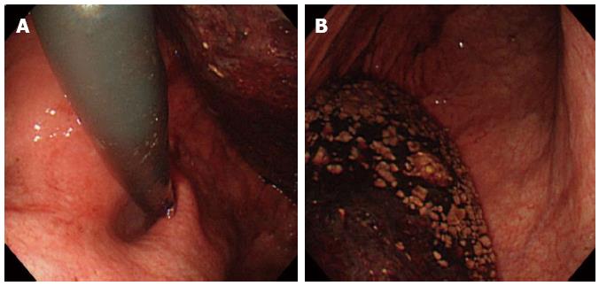
Figure 1 Emergency endoscopic view.
A: The presence of large amounts of blood clots; B: The presence of food residue in the stomach complicated the observation of the region from the gastric fornix to the upper corpus.
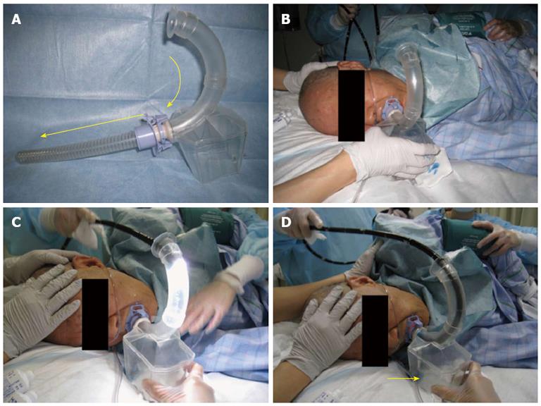
Figure 2 Outer appearance and procedures of using the inverted overtube.
A: Outer appearance of the inverted overtube; B: Switching the patient from the conventional left lateral decubitus position to the right lateral decubitus position using the inverted overtube to dislodge blood clots during emergency endoscopic hemostasis; C: Insertion of the endoscope into the stomach through the inverted overtube in the right lateral decubitus position; D: Continuous aspiration from the bottom of the box (yellow arrow) enabled massive blood clots to be eliminated from the overtube, which maintained a clear endoscopic view during hemostasis.
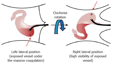
Figure 3 The schema of the visibility differences between 2 positions.
We clockwise rotated the patient’s position to a right lateral decubitus position to dislodge any massive clots and food residue.
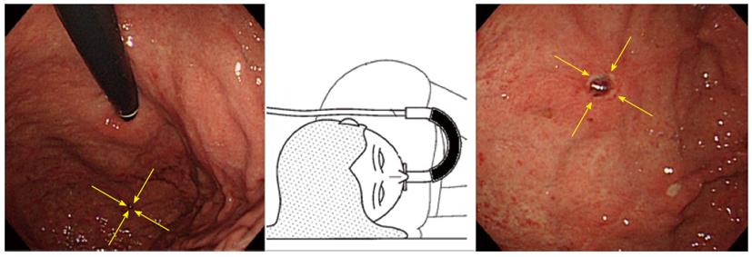
Figure 4 Obtaining a clear view of the gastric fornix and exposed vessels of a Dieulafoy’s ulcer.
Obtaining a clear view of the gastric fornix and upper gastric corpus, with all of the blood clots and food residue dislodged into the gastric antrum and duodenum. The exposed vessels of a Dieulafoy’s ulcer were revealed (yellow arrows). The schema of the inverted overtube is shown in the center.
- Citation: Mori H, Kobara H, Fujihara S, Nishiyama N, Oryu M, Rafiq K, Masaki T. Accurate hemostasis with a new endoscopic overtube for emergency endoscopy. World J Gastroenterol 2013; 19(17): 2723-2726
- URL: https://www.wjgnet.com/1007-9327/full/v19/i17/2723.htm
- DOI: https://dx.doi.org/10.3748/wjg.v19.i17.2723


