Copyright
©2013 Baishideng Publishing Group Co.
World J Gastroenterol. Apr 28, 2013; 19(16): 2543-2549
Published online Apr 28, 2013. doi: 10.3748/wjg.v19.i16.2543
Published online Apr 28, 2013. doi: 10.3748/wjg.v19.i16.2543
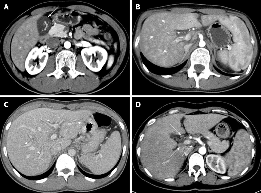
Figure 1 Typical multi-channel computed tomography findings in patients with acute hepatitis.
A: Gallbladder wall thickening (arrow); B: Arterial heterogeneity (asterisks), diffuse heterogeneous attenuation of liver parenchyma in the arterial phase; C: Periportal tracking (arrowheads), decreased attenuation, which highlights the portal vein; D: Lymphadenopathy (arrows) in portal hepatis. Other usual findings, e.g., ascites and splenomegaly, are not shown in this figure.
- Citation: Park SJ, Kim JD, Seo YS, Park BJ, Kim MJ, Um SH, Kim CH, Yim HJ, Baik SK, Jung JY, Keum B, Jeen YT, Lee HS, Chun HJ, Kim CD, Ryu HS. Computed tomography findings for predicting severe acute hepatitis with prolonged cholestasis. World J Gastroenterol 2013; 19(16): 2543-2549
- URL: https://www.wjgnet.com/1007-9327/full/v19/i16/2543.htm
- DOI: https://dx.doi.org/10.3748/wjg.v19.i16.2543


