Copyright
©2013 Baishideng Publishing Group Co.
World J Gastroenterol. Mar 28, 2013; 19(12): 1992-1996
Published online Mar 28, 2013. doi: 10.3748/wjg.v19.i12.1992
Published online Mar 28, 2013. doi: 10.3748/wjg.v19.i12.1992
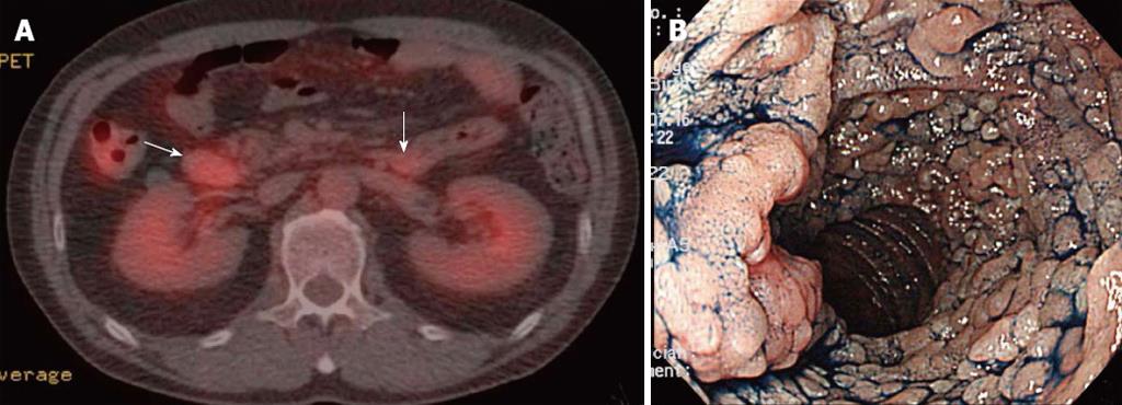
Figure 1 A 52-year-old man with follicular lymphoma of the gastrointestinal tract.
A: 18F-fluorodeoxyglucose positron emission tomography combined with computed tomography in transaxial images showed focal hypermetabolic activities (maximum standardized uptake value, 6.6 and 5.5, respectively) in the lesions of the descending portion of the duodenum and the duodenojejunal flexure (arrows), respectively; B: The esophago-gastro-duodenoscopic view with indigo carmine dye-spray of the descending portion of the duodenum. Numerous whitish granules densely clustered together.
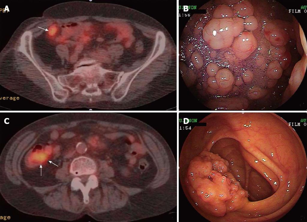
Figure 2 A 66-year-old woman with follicular lymphoma of the gastrointestinal tract.
A, C: 18F-fluorodeoxyglucose positron emission tomography combined with computed tomography in transaxial images showed focal hypermetabolic activities (maximum standardized uptake value, 6.73) in the lesion of the terminal ileum (A) (arrow) and the cecum (C) (arrows); B: The colonoscopic view with indigo carmine dye-spray of the terminal ileum. Numerous polypoid lesions of varying sizes (granules to small nodules) were densely clustered; D: The colonoscopic view of the ileocecal valve. Numerous polypoid lesions of varying sizes (granules to small nodules) were densely clustered.
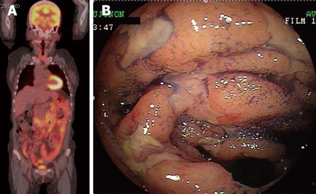
Figure 3 A 61-year-old woman with follicular lymphoma of the gastrointestinal tract.
A: 18F-fluorodeoxyglucose positron emission tomography combined with computed tomography in a projected image showed hypermetabolic foci in broad areas of the gastrointestinal tract from the 2nd portion of the duodenum to the terminal ileum (maximum standardized uptake value, 5.6); B: The colonoscopic view with indigo carmine dye-spray of the terminal ileum. Multiple ulcers with irregular margins were observed.
- Citation: Tari A, Asaoku H, Kunihiro M, Tanaka S, Yoshino T. Usefulness of positron emission tomography in primary intestinal follicular lymphoma. World J Gastroenterol 2013; 19(12): 1992-1996
- URL: https://www.wjgnet.com/1007-9327/full/v19/i12/1992.htm
- DOI: https://dx.doi.org/10.3748/wjg.v19.i12.1992


