©2013 Baishideng Publishing Group Co.
World J Gastroenterol. Mar 21, 2013; 19(11): 1841-1844
Published online Mar 21, 2013. doi: 10.3748/wjg.v19.i11.1841
Published online Mar 21, 2013. doi: 10.3748/wjg.v19.i11.1841
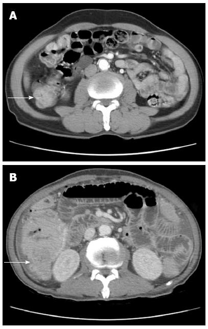
Figure 1 An abdominopelvic computed tomography shows segmental wall thickening of distal ascending with nonspecific multiple small lymphnodes along ileocolic vessels.
Large irregular mass (A) in right ascending colon along hepatic flexure (B).
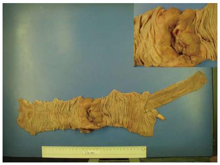
Figure 2 A gross specimen from the ascending colon after right hemicolectomy.
In perforated site, there is an ulcero infiltrative mass measuring 9 cm × 7 cm × 3 cm which is 13 cm away from ileocecal valve and distal resection margin, respectively. The tumor is grayish tan, fish-fresh and appears to extend to serosa.
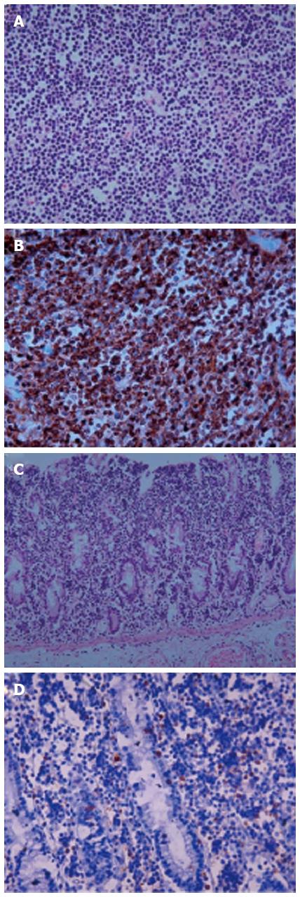
Figure 3 Histologic features in enteropathy-associated T-cell lymphoma type II.
A: The lymphoma cells are small to medium-sized with round, hyperchromatic nuclei (x 400); B: The cells express CD56 (x 400); C, D: The adjacent mucosa shows heavy lymphoid infiltrate (x 200) (C) and CD8 positive intraepithelial lymphocytes (x 400) (D).
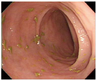
Figure 4 Colonoscopy revealed normal mucosa pattern in whole colonic tract.
- Citation: Kim JB, Kim SH, Cho YK, Ahn SB, Jo YJ, Park YS, Lee JH, Kim DH, Lee H, Jung YY. A case of colon perforation due to enteropathy-associated T-cell lymphoma. World J Gastroenterol 2013; 19(11): 1841-1844
- URL: https://www.wjgnet.com/1007-9327/full/v19/i11/1841.htm
- DOI: https://dx.doi.org/10.3748/wjg.v19.i11.1841


