©2013 Baishideng Publishing Group Co.
World J Gastroenterol. Mar 21, 2013; 19(11): 1820-1826
Published online Mar 21, 2013. doi: 10.3748/wjg.v19.i11.1820
Published online Mar 21, 2013. doi: 10.3748/wjg.v19.i11.1820
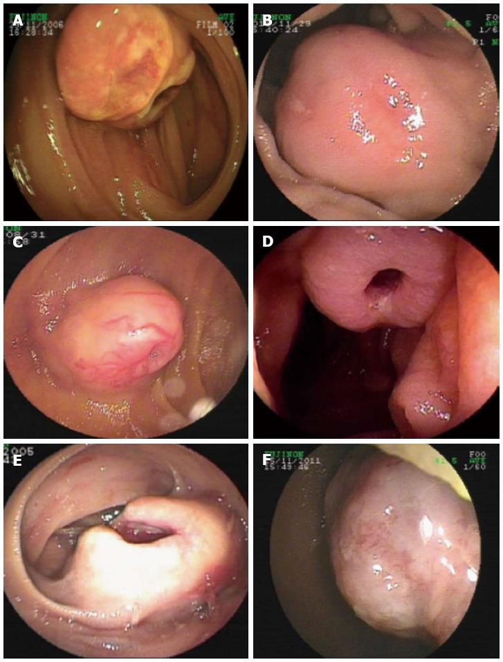
Figure 1 Unsmooth surface of verified gastrointestinal mesenchymal tumors, showing the appearance of erosion or ulcer.
A, B, F: Ulcerative lesions in the surface of the tumors; C, D, E: Ulcerative and depressed pits (A-D: Gastrointestinal mesenchymal tumor; E: Leiomyoma; F: Lipoma).
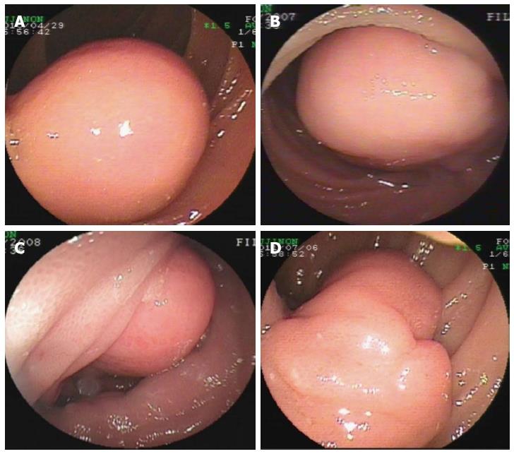
Figure 2 Morphology of verified tumors with smooth surface, indicating tumors with sessile base in round or oval shape.
A-C: Single tumor with round shape and smooth surface (A, B: Gastrointestinal mesenchymal tumor; C: Lipoma; D: A polyp-like tumor with expanded tail, and hemangioma was confirmed by post-surgical pathology).
- Citation: He Q, Bai Y, Zhi FC, Gong W, Gu HX, Xu ZM, Cai JQ, Pan DS, Jiang B. Double-balloon enteroscopy for mesenchymal tumors of small bowel: Nine years’ experience. World J Gastroenterol 2013; 19(11): 1820-1826
- URL: https://www.wjgnet.com/1007-9327/full/v19/i11/1820.htm
- DOI: https://dx.doi.org/10.3748/wjg.v19.i11.1820


