©2013 Baishideng Publishing Group Co.
World J Gastroenterol. Mar 21, 2013; 19(11): 1788-1796
Published online Mar 21, 2013. doi: 10.3748/wjg.v19.i11.1788
Published online Mar 21, 2013. doi: 10.3748/wjg.v19.i11.1788
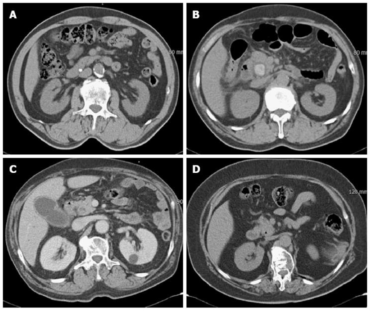
Figure 1 Attenuation patterns of common bile duct stones on multidetector computed tomography.
A: Heavily calcified; B: Radiopaque; C: Less radiopaque; D: Gas attenuation in the stone.
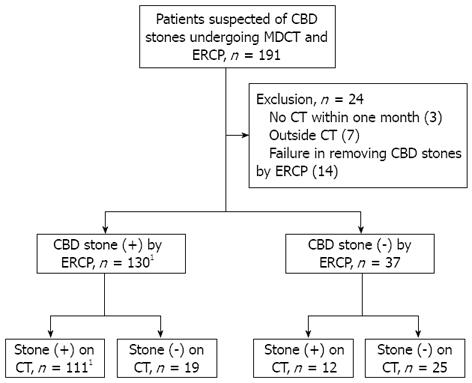
Figure 2 Flow chart of patients with suspicion of common bile duct stones undergoing multidetector computed tomography and endoscopic retrograde cholangiopancreatography.
The sensitivity of multidetector computed tomography (CT) for common bile duct stones was 85.4%. 1Two patients had recurrent common bile duct stones during the study period. CBD: Common bile duct; ERCP: Endoscopic retrograde cholangiopancreatography.
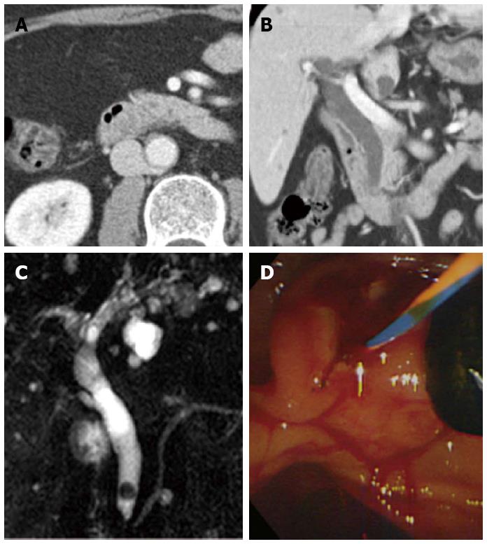
Figure 3 A case of difficult discrimination of common bile duct stone on the axial image of multidetector computed tomography.
The coronal reconstructed image was helpful. A: The stone was ambiguous on the portal venous-phase axial computed tomography (CT) scan; B: The coronal reconstructed CT scan showed a less radiopaque stone near the major ampulla; C, D: Magnetic retrograde cholangiopancreatography and endoscopic retrograde cholangiopancreatography showed a six mm, mixed common bile duct stone.
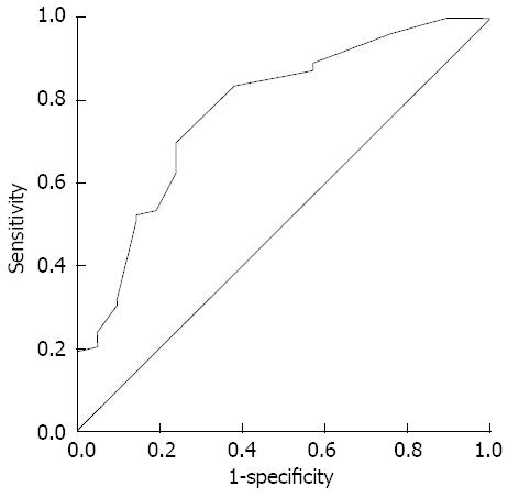
Figure 4 Receiver operating characteristic curve of the detectability of multidetector computed tomography for common bile duct stones according to stone size.
The area under the curve was 0.779 and the optimal cut-off value of stone size was 5 mm.
- Citation: Kim CW, Chang JH, Lim YS, Kim TH, Lee IS, Han SW. Common bile duct stones on multidetector computed tomography: Attenuation patterns and detectability. World J Gastroenterol 2013; 19(11): 1788-1796
- URL: https://www.wjgnet.com/1007-9327/full/v19/i11/1788.htm
- DOI: https://dx.doi.org/10.3748/wjg.v19.i11.1788


